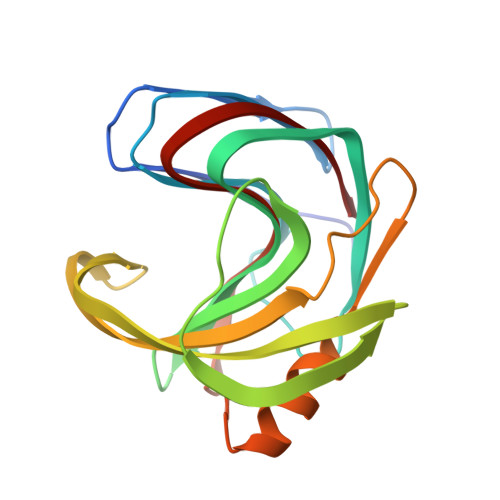
5TVV

Computationally Designed Fentanyl Binder - Fen49* Apo


Experimental Data Snapshot

  • Method: X-RAY DIFFRACTION
  • Resolution: 1.79 Å
  • R-Value Free: 0.225 
  • R-Value Work: 0.204 
  • R-Value Observed: 0.205 

Starting Model: experimental
View more details

wwPDB Validation   3D Report Full Report


This is version 1.3 of the entry. See complete history


Literature

Computational design of environmental sensors for the potent opioid fentanyl.

Bick, M.J.Greisen, P.J.Morey, K.J.Antunes, M.S.La, D.Sankaran, B.Reymond, L.Johnsson, K.Medford, J.I.Baker, D.

(2017) Elife 6

  • DOI: https://doi.org/10.7554/eLife.28909
  • Primary Citation of Related Structures:  
    5TVV, 5TVY, 5TZO

  • PubMed Abstract: 

    We describe the computational design of proteins that bind the potent analgesic fentanyl. Our approach employs a fast docking algorithm to find shape complementary ligand placement in protein scaffolds, followed by design of the surrounding residues to optimize binding affinity. Co-crystal structures of the highest affinity binder reveal a highly preorganized binding site, and an overall architecture and ligand placement in close agreement with the design model. We use the designs to generate plant sensors for fentanyl by coupling ligand binding to design stability. The method should be generally useful for detecting toxic hydrophobic compounds in the environment.


  • Organizational Affiliation

    Department of Biochemistry, University of Washington, Seattle, United States.


Macromolecules
Find similar proteins by:  (by identity cutoff)  |  3D Structure
Entity ID: 1
MoleculeChains Sequence LengthOrganismDetailsImage
Endo-1,4-beta-xylanase A
A, B, C
188Bacillus subtilis subsp. subtilis str. 168Mutation(s): 11 
Gene Names: xynABSU18840
EC: 3.2.1.8
UniProt
Find proteins for P18429 (Bacillus subtilis (strain 168))
Explore P18429 
Go to UniProtKB:  P18429
Entity Groups  
Sequence Clusters30% Identity50% Identity70% Identity90% Identity95% Identity100% Identity
UniProt GroupP18429
Sequence Annotations
Expand
  • Reference Sequence
Experimental Data & Validation

Experimental Data

  • Method: X-RAY DIFFRACTION
  • Resolution: 1.79 Å
  • R-Value Free: 0.225 
  • R-Value Work: 0.204 
  • R-Value Observed: 0.205 
  • Space Group: P 21 21 21
Unit Cell:
Length ( Å )Angle ( ˚ )
a = 54.76α = 90
b = 73.259β = 90
c = 137.558γ = 90
Software Package:
Software NamePurpose
HKL-2000data reduction
HKL-2000data scaling
PHASERphasing
Cootmodel building
PHENIXrefinement
PDB_EXTRACTdata extraction
HKLdata scaling

Structure Validation

View Full Validation Report


Entry History & Funding Information

Deposition Data


Funding OrganizationLocationGrant Number
National Institutes of Health/National Cancer Institute (NIH/NCI)United States5F32CA171572-03
Defense Threat Reduction Agency (DTRA)United StatesHDTRA1-11-1-0041
Life Sciences Discovery FundUnited States9598385

Revision History  (Full details and data files)

  • Version 1.0: 2017-10-04
    Type: Initial release
  • Version 1.1: 2019-12-04
    Changes: Author supporting evidence
  • Version 1.2: 2020-01-01
    Changes: Author supporting evidence
  • Version 1.3: 2023-10-04
    Changes: Data collection, Database references, Derived calculations, Refinement description