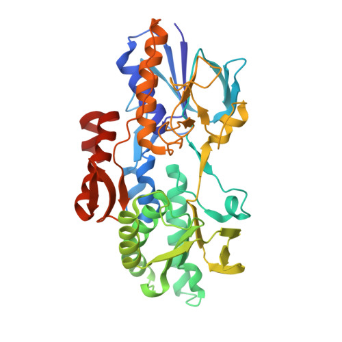
5WED | pdb_00005wed

Structure of bacterial type II NADH dehydrogenase from Caldalkalibacillus thermarum at 2.15A resolution


Experimental Data Snapshot

  • Method: X-RAY DIFFRACTION
  • Resolution: 2.15 Å
  • R-Value Free: 
    0.238 (Depositor), 0.240 (DCC) 
  • R-Value Work: 
    0.208 (Depositor), 0.210 (DCC) 
  • R-Value Observed: 
    0.209 (Depositor) 

wwPDB Validation   3D Report Full Report


Ligand Structure Quality Assessment 

Created with Raphaël 2.3.0Worse 01 BetterLigand structure goodness of fit to experimental dataBest fitted FADClick on this verticalbar to view details

This is version 1.4 of the entry. See complete history


Literature

Crystal structure of type II NADH:quinone oxidoreductase from Caldalkalibacillus thermarum with an improved resolution of 2.15 angstrom.

Nakatani, Y.Jiao, W.Aragao, D.Shimaki, Y.Petri, J.Parker, E.J.Cook, G.M.

(2017) Acta Crystallogr F Struct Biol Commun 73: 541-549

  • DOI: https://doi.org/10.1107/S2053230X17013073
  • Primary Citation of Related Structures:  
    5WED

  • PubMed Abstract: 

    Type II NADH:quinone oxidoreductase (NDH-2) is a respiratory enzyme found in the electron-transport chain of many species, with the exception of mammals. It is a 40-70 kDa single-subunit monotopic membrane protein that catalyses the oxidation of NADH and the reduction of quinone molecules via the cofactor FAD. NDH-2 is a promising new target for drug development given its essential role in many bacterial species and intracellular parasites. Only two bacterial NDH-2 structures have been reported and these structures are at moderate resolution (2.3-2.5 Å). In this communication, a new crystallization platform is reported that produced high-quality NDH-2 crystals that diffracted to high resolution (2.15 Å). The high-resolution NDH-2 structure was used for in silico quinone substrate-docking studies to investigate the binding poses of menadione and ubiquinone molecules. These studies revealed that a very limited number of molecular interactions occur at the quinone-binding site of NDH-2. Given that the conformation of the active site is well defined, this high-resolution structure is potentially suitable for in silico inhibitor-compound screening and ligand-docking applications.


  • Organizational Affiliation

    Department of Microbiology and Immunology, University of Otago, 720 Cumberland Street, Dunedin 9054, New Zealand.


Macromolecules
Find similar proteins by:  (by identity cutoff)  |  3D Structure
Entity ID: 1
MoleculeChains Sequence LengthOrganismDetailsImage
FAD-dependent pyridine nucleotide-disulfide oxidoreductaseA [auth B],
B [auth A],
C,
D
405Caldalkalibacillus thermarum TA2.A1Mutation(s): 0 
Gene Names: CathTA2_0279
UniProt
Find proteins for F5L3B8 (Caldalkalibacillus thermarum (strain TA2.A1))
Explore F5L3B8 
Go to UniProtKB:  F5L3B8
Entity Groups  
Sequence Clusters30% Identity50% Identity70% Identity90% Identity95% Identity100% Identity
UniProt GroupF5L3B8
Sequence Annotations
Expand
  • Reference Sequence
Experimental Data & Validation

Experimental Data

  • Method: X-RAY DIFFRACTION
  • Resolution: 2.15 Å
  • R-Value Free:  0.238 (Depositor), 0.240 (DCC) 
  • R-Value Work:  0.208 (Depositor), 0.210 (DCC) 
  • R-Value Observed: 0.209 (Depositor) 
Space Group: P 1 21 1
Unit Cell:
Length ( Å )Angle ( ˚ )
a = 72.804α = 90
b = 113.57β = 91.02
c = 129.781γ = 90
Software Package:
Software NamePurpose
PHENIXrefinement
XDSdata reduction
Aimlessdata scaling
PHASERphasing

Structure Validation

View Full Validation Report


Ligand Structure Quality Assessment 

Created with Raphaël 2.3.0Worse 01 BetterLigand structure goodness of fit to experimental dataBest fitted FADClick on this verticalbar to view details

Entry History & Funding Information

Deposition Data


Funding OrganizationLocationGrant Number
Health Research Council (HRC)New Zealand--
Maurice Wilkins Centre for Molecular BiodiscoveryNew Zealand--

Revision History  (Full details and data files)

  • Version 1.0: 2017-10-18
    Type: Initial release
  • Version 1.1: 2018-04-18
    Changes: Data collection, Database references
  • Version 1.2: 2020-01-01
    Changes: Author supporting evidence
  • Version 1.3: 2023-03-29
    Changes: Database references, Structure summary
  • Version 1.4: 2024-05-22
    Changes: Data collection