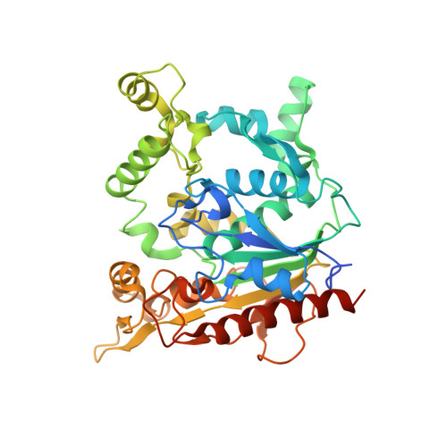
6KSL

Staphylococcus aureus lipase - S116A inactive mutant


Experimental Data Snapshot

  • Method: X-RAY DIFFRACTION
  • Resolution: 2.59 Å
  • R-Value Free: 0.268 
  • R-Value Work: 0.249 
  • R-Value Observed: 0.250 

Starting Model: experimental
View more details

wwPDB Validation   3D Report Full Report


Ligand Structure Quality Assessment 


This is version 1.1 of the entry. See complete history


Literature

Crystal structure of pathogenic Staphylococcus aureus lipase complex with the anti-obesity drug orlistat.

Kitadokoro, K.Tanaka, M.Hikima, T.Okuno, Y.Yamamoto, M.Kamitani, S.

(2020) Sci Rep 10: 5469-5469

  • DOI: https://doi.org/10.1038/s41598-020-62427-8
  • Primary Citation of Related Structures:  
    6KSI, 6KSL, 6KSM

  • PubMed Abstract: 

    Staphylococcus aureus lipase (SAL), a triacylglycerol esterase, is an important virulence factor and may be a therapeutic target for infectious diseases. Herein, we determined the 3D structure of native SAL, the mutated S116A inactive form, and the inhibitor complex using the anti-obesity drug orlistat to aid in drug development. The determined crystal structures showed a typical α/β hydrolase motif with a dimeric form. Fatty acids bound near the active site in native SAL and inactive S116A mutant structures. We found that orlistat potently inhibits SAL activity, and it covalently bound to the catalytic Ser116 residue. This is the first report detailing orlistat-lipase binding. It provides structure-based information on the production of potent anti-SAL drugs and lipase inhibitors. These results also indicated that orlistat can be repositioned to treat bacterial diseases.


  • Organizational Affiliation

    Faculty of Molecular Chemistry and Engineering, Graduate School of Science and Technology, Kyoto Institute of Technology, Hashigami-cho, Matsugasaki, Sakyo-ku, Kyoto, 606-8585, Japan. kengo@kit.ac.jp.


Macromolecules
Find similar proteins by:  (by identity cutoff)  |  3D Structure
Entity ID: 1
MoleculeChains Sequence LengthOrganismDetailsImage
Lipase 2
A, B
408Staphylococcus aureusMutation(s): 2 
Gene Names: lipBN1321_80040
EC: 3.1.1.3
UniProt
Find proteins for A0A0U1MWF9 (Staphylococcus aureus)
Explore A0A0U1MWF9 
Go to UniProtKB:  A0A0U1MWF9
Entity Groups  
Sequence Clusters30% Identity50% Identity70% Identity90% Identity95% Identity100% Identity
UniProt GroupA0A0U1MWF9
Sequence Annotations
Expand
  • Reference Sequence
Small Molecules
Ligands 4 Unique
IDChains Name / Formula / InChI Key2D Diagram3D Interactions
DAO (Subject of Investigation/LOI)
Query on DAO

Download Ideal Coordinates CCD File 
E [auth A],
I [auth B],
J [auth B]
LAURIC ACID
C12 H24 O2
POULHZVOKOAJMA-UHFFFAOYSA-N
BUA (Subject of Investigation/LOI)
Query on BUA

Download Ideal Coordinates CCD File 
F [auth A],
K [auth B]
butanoic acid
C4 H8 O2
FERIUCNNQQJTOY-UHFFFAOYSA-N
ZN (Subject of Investigation/LOI)
Query on ZN

Download Ideal Coordinates CCD File 
C [auth A],
G [auth B]
ZINC ION
Zn
PTFCDOFLOPIGGS-UHFFFAOYSA-N
CA (Subject of Investigation/LOI)
Query on CA

Download Ideal Coordinates CCD File 
D [auth A],
H [auth B]
CALCIUM ION
Ca
BHPQYMZQTOCNFJ-UHFFFAOYSA-N
Experimental Data & Validation

Experimental Data

  • Method: X-RAY DIFFRACTION
  • Resolution: 2.59 Å
  • R-Value Free: 0.268 
  • R-Value Work: 0.249 
  • R-Value Observed: 0.250 
  • Space Group: P 61 2 2
Unit Cell:
Length ( Å )Angle ( ˚ )
a = 164.209α = 90
b = 164.209β = 90
c = 233.053γ = 120
Software Package:
Software NamePurpose
REFMACrefinement
PDB_EXTRACTdata extraction
XDSdata reduction
SCALAdata scaling
PHASERphasing

Structure Validation

View Full Validation Report


Ligand Structure Quality Assessment 


Entry History 

Deposition Data

Revision History  (Full details and data files)

  • Version 1.0: 2020-04-08
    Type: Initial release
  • Version 1.1: 2023-11-22
    Changes: Data collection, Database references, Derived calculations, Refinement description, Structure summary