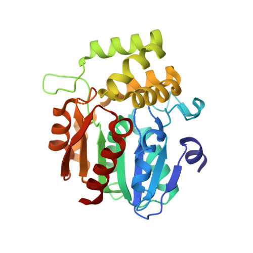
6O5J | pdb_00006o5j

Crystal Structure of DAD2 bound to quinazolinone derivative


Experimental Data Snapshot

  • Method: X-RAY DIFFRACTION
  • Resolution: 1.63 Å
  • R-Value Free: 
    0.182 (Depositor), 0.190 (DCC) 
  • R-Value Work: 
    0.144 (Depositor), 0.150 (DCC) 
  • R-Value Observed: 
    0.146 (Depositor) 

Starting Model: experimental
View more details

wwPDB Validation   3D Report Full Report


Ligand Structure Quality Assessment 

Created with Raphaël 2.3.0Worse 01 BetterLigand structure goodness of fit to experimental dataBest fitted LM7Click on this verticalbar to view details

This is version 1.2 of the entry. See complete history


Literature

Chemical synthesis and characterization of a new quinazolinedione competitive antagonist for strigolactone receptors with an unexpected binding mode.

Hamiaux, C.Larsen, L.Lee, H.W.Luo, Z.Sharma, P.Hawkins, B.C.Perry, N.B.Snowden, K.C.

(2019) Biochem J 476: 1843-1856

  • DOI: https://doi.org/10.1042/BCJ20190288
  • Primary Citation of Related Structures:  
    6O5J

  • PubMed Abstract: 

    Strigolactones (SLs) are multifunctional plant hormones regulating essential physiological processes affecting growth and development. In vascular plants, SLs are recognized by α/β hydrolase-fold proteins from the D14/DAD2 (Dwarf14/Decreased Apical Dominance 2) family in the initial step of the signaling pathway. We have previously discovered that N -phenylanthranilic acid derivatives (e.g. tolfenamic acid) are potent antagonists of SL receptors, prompting us to design quinazolinone and quinazolinedione derivatives (QADs and QADDs, respectively) as second-generation antagonists. Initial in silico docking studies suggested that these compounds would bind to DAD2, the petunia SL receptor, with higher affinity than the first-generation compounds. However, only one of the QADs/QADDs tested in in vitro assays acted as a competitive antagonist of SL receptors, with reduced affinity and potency compared with its N -phenylanthranilic acid 'parent'. X-ray crystal structure analysis revealed that the binding mode of the active QADD inside DAD2's cavity was not that predicted in silico , highlighting a novel inhibition mechanism for SL receptors. Despite a ∼10-fold difference in potency in vitro , the QADD and tolfenamic acid had comparable activity in planta , suggesting that the QADD compensates for lower potency with increased bioavailability. Altogether, our results establish this QADD as a novel lead compound towards the development of potent and bioavailable antagonists of SL receptors.


  • Organizational Affiliation

    The New Zealand Institute for Plant and Food Research Limited, Auckland, New Zealand cyril.hamiaux@plantandfood.co.nz.


Macromolecules
Find similar proteins by:  (by identity cutoff)  |  3D Structure
Entity ID: 1
MoleculeChains Sequence LengthOrganismDetailsImage
Probable strigolactone esterase DAD2
A, B
264Petunia x hybridaMutation(s): 1 
Gene Names: DAD2
EC: 3.1
UniProt
Find proteins for J9U5U9 (Petunia hybrida)
Explore J9U5U9 
Go to UniProtKB:  J9U5U9
Entity Groups  
Sequence Clusters30% Identity50% Identity70% Identity90% Identity95% Identity100% Identity
UniProt GroupJ9U5U9
Sequence Annotations
Expand
  • Reference Sequence
Small Molecules
Ligands 4 Unique
IDChains Name / Formula / InChI Key2D Diagram3D Interactions
LM7
Query on LM7

Download Ideal Coordinates CCD File 
C [auth A],
F [auth B]
1-(4-hydroxy-3-nitrophenyl)quinazoline-2,4(1H,3H)-dione
C14 H9 N3 O5
YVQCKIRAVMSRGN-UHFFFAOYSA-N
PEG
Query on PEG

Download Ideal Coordinates CCD File 
G [auth B]DI(HYDROXYETHYL)ETHER
C4 H10 O3
MTHSVFCYNBDYFN-UHFFFAOYSA-N
GOL
Query on GOL

Download Ideal Coordinates CCD File 
D [auth A],
H [auth B]
GLYCEROL
C3 H8 O3
PEDCQBHIVMGVHV-UHFFFAOYSA-N
ACT
Query on ACT

Download Ideal Coordinates CCD File 
E [auth A]ACETATE ION
C2 H3 O2
QTBSBXVTEAMEQO-UHFFFAOYSA-M
Experimental Data & Validation

Experimental Data

  • Method: X-RAY DIFFRACTION
  • Resolution: 1.63 Å
  • R-Value Free:  0.182 (Depositor), 0.190 (DCC) 
  • R-Value Work:  0.144 (Depositor), 0.150 (DCC) 
  • R-Value Observed: 0.146 (Depositor) 
Space Group: P 1
Unit Cell:
Length ( Å )Angle ( ˚ )
a = 36.63α = 95.69
b = 56.83β = 94.59
c = 68.8γ = 108.74
Software Package:
Software NamePurpose
REFMACrefinement
MOSFLMdata reduction
Aimlessdata scaling
PHASERphasing
PDB_EXTRACTdata extraction

Structure Validation

View Full Validation Report


Ligand Structure Quality Assessment 

Created with Raphaël 2.3.0Worse 01 BetterLigand structure goodness of fit to experimental dataBest fitted LM7Click on this verticalbar to view details

Entry History & Funding Information

Deposition Data

  • Released Date: 2019-06-26 
  • Deposition Author(s): Hamiaux, C.

Funding OrganizationLocationGrant Number
Royal Society of New ZealandNew ZealandPAF1301

Revision History  (Full details and data files)

  • Version 1.0: 2019-06-26
    Type: Initial release
  • Version 1.1: 2019-07-10
    Changes: Data collection, Database references
  • Version 1.2: 2023-10-11
    Changes: Data collection, Database references, Refinement description