


Crystal structure of Plasmodium falciparum PdxK with ligands AMP-PNP and PL.
Gao, K., Wang, W., Groves, M.R.(2019) Crystals (Basel)
Experimental Data Snapshot
Starting Model: experimental
View more details
(2019) Crystals (Basel)
Entity ID: 1 | |||||
|---|---|---|---|---|---|
| Molecule | Chains | Sequence Length | Organism | Details | Image |
| Pyridoxal kinase | 497 | Plasmodium falciparum 3D7 | Mutation(s): 0 Gene Names: PF3D7_0616000 EC: 2.7.1.35 | 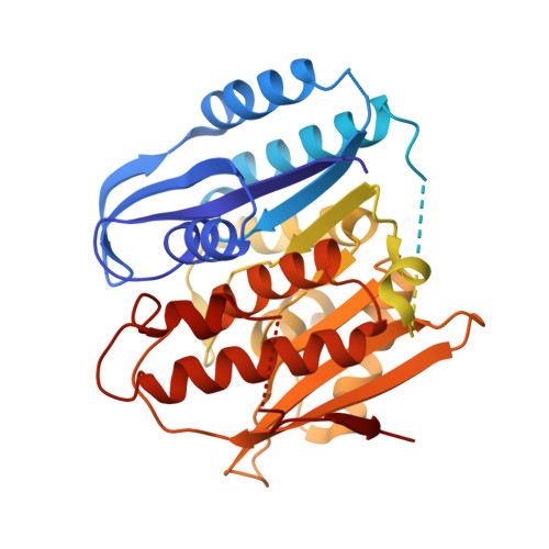 | |
UniProt | |||||
Find proteins for C6KT01 (Plasmodium falciparum (isolate 3D7)) Explore C6KT01 Go to UniProtKB: C6KT01 | |||||
Entity Groups | |||||
| Sequence Clusters | 30% Identity50% Identity70% Identity90% Identity95% Identity100% Identity | ||||
| UniProt Group | C6KT01 | ||||
Sequence AnnotationsExpand | |||||
| |||||
| Ligands 3 Unique | |||||
|---|---|---|---|---|---|
| ID | Chains | Name / Formula / InChI Key | 2D Diagram | 3D Interactions | |
| ANP Query on ANP | C [auth A], G [auth B] | PHOSPHOAMINOPHOSPHONIC ACID-ADENYLATE ESTER C10 H17 N6 O12 P3 PVKSNHVPLWYQGJ-KQYNXXCUSA-N |  | ||
| PXL Query on PXL | D [auth A], H [auth B] | 3-HYDROXY-5-(HYDROXYMETHYL)-2-METHYLISONICOTINALDEHYDE C8 H9 N O3 RADKZDMFGJYCBB-UHFFFAOYSA-N |  | ||
| MG Query on MG | E [auth A], F [auth A], I [auth B] | MAGNESIUM ION Mg JLVVSXFLKOJNIY-UHFFFAOYSA-N |  | ||
| Length ( Å ) | Angle ( ˚ ) |
|---|---|
| a = 52.703 | α = 90 |
| b = 62.004 | β = 94.99 |
| c = 93.712 | γ = 90 |
| Software Name | Purpose |
|---|---|
| PHENIX | refinement |
| PDB_EXTRACT | data extraction |
| XDS | data reduction |
| Aimless | data scaling |
| PHENIX | phasing |


