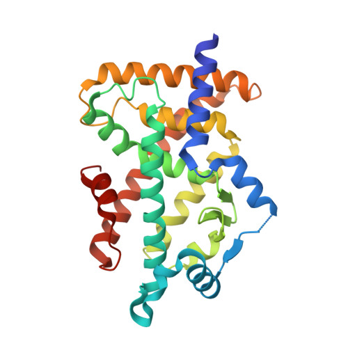
7E5F

HUMAN PPAR ALPHA LIGAND BINDING DOMAIN IN COMPLEX WITH TIPP703 OBTAINED BY SOAKING


Experimental Data Snapshot

  • Method: X-RAY DIFFRACTION
  • Resolution: 1.79 Å
  • R-Value Free: 0.229 
  • R-Value Work: 0.196 
  • R-Value Observed: 0.198 

Starting Model: experimental
View more details

wwPDB Validation   3D Report Full Report


Ligand Structure Quality Assessment 


This is version 1.1 of the entry. See complete history


Literature

Crystal Structures of the Human Peroxisome Proliferator-Activated Receptor (PPAR) alpha Ligand-Binding Domain in Complexes with a Series of Phenylpropanoic Acid Derivatives Generated by a Ligand-Exchange Soaking Method.

Oyama, T.Kamata, S.Ishii, I.Miyachi, H.

(2021) Biol Pharm Bull 44: 1202-1209

  • DOI: https://doi.org/10.1248/bpb.b21-00220
  • Primary Citation of Related Structures:  
    7E5F, 7E5G, 7E5H, 7E5I

  • PubMed Abstract: 

    Peroxisome proliferator-activated receptor (PPAR)α, a member of the nuclear receptor family, is a transcription factor that regulates the expression of genes related to lipid metabolism in a ligand-dependent manner, and has attracted attention as a target for hypolipidemic drugs. We have been developing phenylpropaonic acid derivatives as PPARα-targeted drug candidates for the treatment of metabolic diseases. Recently, we have developed the "ligand-exchange soaking method," which crystallizes the recombinant PPARα ligand-binding domain (LBD) as a complex with intrinsic fatty acids derived from an expression host Escherichia (E.) coli and thereafter replaces them with other higher-affinity ligands by soaking. Here we applied this method for preparation of cocrystals of PPARα LBD with its ligands that have not been obtained with the conventional cocrystallization method. We revealed the high-resolution structures of the cocrystals of PPARα LBD and the three synthetic phenylpropaonic acid derivatives: TIPP-703, APHM19, and YN4pai, the latter two of which are the first observations. The overall structures of cocrystals obtained from the two methods are identical and illustrate the close interaction between these ligands and the surrounding amino acid residues of PPARα LBD. This ligand-exchange soaking method could be applicable to high throughput preparations of co-crystals with another subtype PPARδ LBD for high resolution X-ray crystallography, because it also crystallizes in complex with intrinsic fatty acid(s) while not in the apo-form.


  • Organizational Affiliation

    Faculty of Life and Environmental Sciences, University of Yamanashi.


Macromolecules
Find similar proteins by:  (by identity cutoff)  |  3D Structure
Entity ID: 1
MoleculeChains Sequence LengthOrganismDetailsImage
Peroxisome proliferator-activated receptor alpha273Homo sapiensMutation(s): 0 
UniProt & NIH Common Fund Data Resources
Find proteins for Q07869 (Homo sapiens)
Explore Q07869 
Go to UniProtKB:  Q07869
PHAROS:  Q07869
GTEx:  ENSG00000186951 
Entity Groups  
Sequence Clusters30% Identity50% Identity70% Identity90% Identity95% Identity100% Identity
UniProt GroupQ07869
Sequence Annotations
Expand
  • Reference Sequence
Small Molecules
Ligands 1 Unique
IDChains Name / Formula / InChI Key2D Diagram3D Interactions
S44 (Subject of Investigation/LOI)
Query on S44

Download Ideal Coordinates CCD File 
B [auth A](2S)-2-(4-propoxy-3-{[({4-[(3S,5S,7S)-tricyclo[3.3.1.1~3,7~]dec-1-yl]phenyl}carbonyl)amino]methyl}benzyl)butanoic acid
C32 H41 N O4
GUFHDBFHNDUDJA-ZQPQWVRXSA-N
Binding Affinity Annotations 
IDSourceBinding Affinity
S44 BindingDB:  7E5F EC50: min: 54, max: 140 (nM) from 4 assay(s)
Experimental Data & Validation

Experimental Data

  • Method: X-RAY DIFFRACTION
  • Resolution: 1.79 Å
  • R-Value Free: 0.229 
  • R-Value Work: 0.196 
  • R-Value Observed: 0.198 
  • Space Group: P 1 21 1
Unit Cell:
Length ( Å )Angle ( ˚ )
a = 44.893α = 90
b = 62.158β = 105.782
c = 53.369γ = 90
Software Package:
Software NamePurpose
PHENIXrefinement
XDSdata reduction
Aimlessdata scaling
PHASERphasing

Structure Validation

View Full Validation Report


Ligand Structure Quality Assessment 


Entry History 

Deposition Data

Revision History  (Full details and data files)

  • Version 1.0: 2021-10-06
    Type: Initial release
  • Version 1.1: 2023-11-29
    Changes: Data collection, Refinement description