8ENW | pdb_00008enw

Crystal structure of beta'-COPI-WD40 domain in complex with SARS-CoV-2 clientized spike tail heptapeptide.


Experimental Data Snapshot

  • Method: X-RAY DIFFRACTION
  • Resolution: 1.45 Å
  • R-Value Free: 
    0.180 (Depositor), 0.180 (DCC) 
  • R-Value Work: 
    0.153 (Depositor), 0.150 (DCC) 
  • R-Value Observed: 
    0.155 (Depositor) 

wwPDB Validation   3D Report Full Report


Ligand Structure Quality Assessment 

Created with Raphaël 2.3.0Worse 01 BetterLigand structure goodness of fit to experimental dataBest fitted EDOClick on this verticalbar to view details

This is version 1.0 of the entry. See complete history


Literature

A single C-terminal residue controls SARS-CoV-2 spike trafficking and incorporation into VLPs.

Dey, D.Qing, E.He, Y.Chen, Y.Jennings, B.Cohn, W.Singh, S.Gakhar, L.Schnicker, N.J.Pierce, B.G.Whitelegge, J.P.Doray, B.Orban, J.Gallagher, T.Hasan, S.S.

(2023) Nat Commun 14: 8358-8358

  • DOI: https://doi.org/10.1038/s41467-023-44076-3
  • Primary Citation of Related Structures:  
    8ENS, 8ENW, 8ENX, 8ENY, 8ENZ, 8EO0, 8SZX

  • PubMed Abstract: 

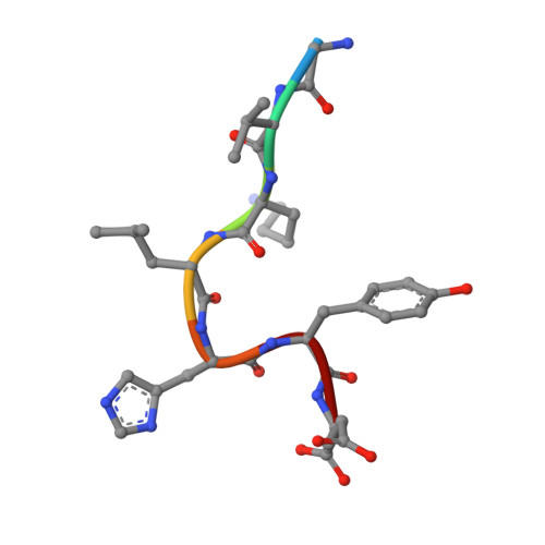
    The spike (S) protein of SARS-CoV-2 is delivered to the virion assembly site in the ER-Golgi Intermediate Compartment (ERGIC) from both the ER and cis-Golgi in infected cells. However, the relevance and modulatory mechanism of this bidirectional trafficking are unclear. Here, using structure-function analyses, we show that S incorporation into virus-like particles (VLP) and VLP fusogenicity are determined by coatomer-dependent S delivery from the cis-Golgi and restricted by S-coatomer dissociation. Although S mimicry of the host coatomer-binding dibasic motif ensures retrograde trafficking to the ERGIC, avoidance of the host-like C-terminal acidic residue is critical for S-coatomer dissociation and therefore incorporation into virions or export for cell-cell fusion. Because this C-terminal residue is the key determinant of SARS-CoV-2 assembly and fusogenicity, our work provides a framework for the export of S protein encoded in genetic vaccines for surface display and immune activation.


  • Organizational Affiliation

    Department of Biochemistry and Molecular Biology, University of Maryland School of Medicine, Baltimore, MD, 21201, USA.


Macromolecules
Find similar proteins by:  (by identity cutoff)  |  3D Structure
Entity ID: 1
MoleculeChains Sequence LengthOrganismDetailsImage
Coatomer subunit beta'
A, B
302Saccharomyces cerevisiaeMutation(s): 0 
UniProt
Find proteins for P41811 (Saccharomyces cerevisiae (strain ATCC 204508 / S288c))
Explore P41811 
Go to UniProtKB:  P41811
Entity Groups  
Sequence Clusters30% Identity50% Identity70% Identity90% Identity95% Identity100% Identity
UniProt GroupP41811
Sequence Annotations
Expand
  • Reference Sequence

Find similar proteins by:  Sequence   |   3D Structure  

Entity ID: 2
MoleculeChains Sequence LengthOrganismDetailsImage
Clientized spike tail peptide
C, D
7Severe acute respiratory syndrome coronavirus 2Mutation(s): 0 
UniProt
Find proteins for P0DTC2 (Severe acute respiratory syndrome coronavirus 2)
Explore P0DTC2 
Go to UniProtKB:  P0DTC2
Entity Groups  
UniProt GroupP0DTC2
Sequence Annotations
Expand
  • Reference Sequence
Small Molecules
Ligands 1 Unique
IDChains Name / Formula / InChI Key2D Diagram3D Interactions
EDO (Subject of Investigation/LOI)
Query on EDO

Download Ideal Coordinates CCD File 
E [auth A]
F [auth A]
G [auth A]
H [auth A]
I [auth A]
1,2-ETHANEDIOL
C2 H6 O2
LYCAIKOWRPUZTN-UHFFFAOYSA-N
Experimental Data & Validation

Experimental Data

  • Method: X-RAY DIFFRACTION
  • Resolution: 1.45 Å
  • R-Value Free:  0.180 (Depositor), 0.180 (DCC) 
  • R-Value Work:  0.153 (Depositor), 0.150 (DCC) 
  • R-Value Observed: 0.155 (Depositor) 
Space Group: P 1
Unit Cell:
Length ( Å )Angle ( ˚ )
a = 42.711α = 81.24
b = 46.012β = 81.52
c = 84.319γ = 69.01
Software Package:
Software NamePurpose
PHENIXrefinement
HKL-3000data reduction
XDSdata scaling
PHASERphasing

Structure Validation

View Full Validation Report


Ligand Structure Quality Assessment 

Created with Raphaël 2.3.0Worse 01 BetterLigand structure goodness of fit to experimental dataBest fitted EDOClick on this verticalbar to view details

Entry History & Funding Information

Deposition Data


Funding OrganizationLocationGrant Number
National Institutes of Health/National Cancer Institute (NIH/NCI)United StatesP30CA134274
National Institutes of Health/National Institute of General Medical Sciences (NIH/NIGMS)United StatesR01GM150187

Revision History  (Full details and data files)

  • Version 1.0: 2024-01-31
    Type: Initial release