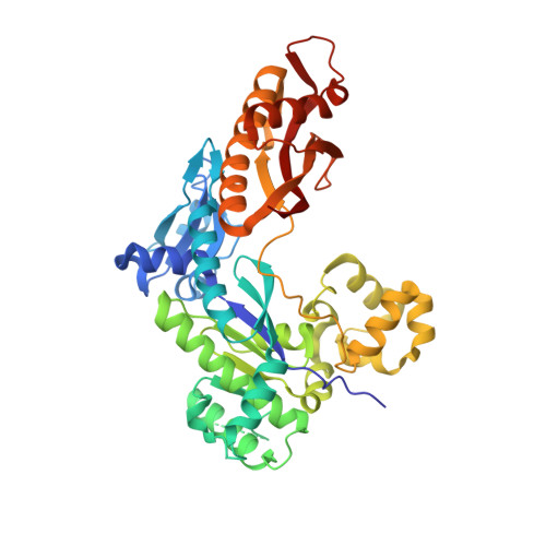
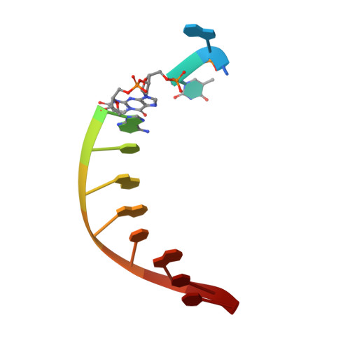
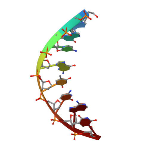
8EVE

HUMAN DNA POLYMERASE ETA INSERTION COMPLEX


Experimental Data Snapshot

  • Method: X-RAY DIFFRACTION
  • Resolution: 2.35 Å
  • R-Value Free: 0.244 
  • R-Value Work: 0.189 
  • R-Value Observed: 0.192 

wwPDB Validation   3D Report Full Report


This is version 1.1 of the entry. See complete history


Literature

The peroxidation-derived DNA adduct, 6-oxo-M 1 dG, is a strong block to replication by human DNA polymerase eta.

Richie-Jannetta, R.Pallan, P.Kingsley, P.J.Kamdar, N.Egli, M.Marnett, L.J.

(2023) J Biol Chem 299: 105067-105067

  • DOI: https://doi.org/10.1016/j.jbc.2023.105067
  • Primary Citation of Related Structures:  
    8EVE, 8EVF

  • PubMed Abstract: 

    The DNA adduct 6-oxo-M 1 dG, (3-(2'-deoxy-β-D-erythro-pentofuranosyl)-6-oxo-pyrimido(1,2alpha)purin-10(3H)-one) is formed in the genome via oxidation of the peroxidation-derived adduct M 1 dG. However, the effect of 6-oxo-M 1 dG adducts on subsequent DNA replication is unclear. Here we investigated the ability of the human Y-family polymerase hPol η to bypass 6-oxo-M 1 dG. Using steady-state kinetics and analysis of DNA extension products by liquid chromatography-tandem mass spectrometry, we found hPol η preferentially inserts a dAMP or dGMP nucleotide into primer-templates across from the 6-oxo-M 1 dG adduct, with dGMP being slightly preferred. We also show primer-templates with a 3'-terminal dGMP or dAMP across from 6-oxo-M 1 dG were extended to a greater degree than primers with a dCMP or dTMP across from the adduct. In addition, we explored the structural basis for bypass of 6-oxo-M 1 dG by hPol η using X-ray crystallography of both an insertion-stage and an extension-stage complex. In the insertion-stage complex, we observed that the incoming dCTP opposite 6-oxo-M 1 dG, although present during crystallization, was not present in the active site. We found the adduct does not interact with residues in the hPol η active site but rather forms stacking interactions with the base pair immediately 3' to the adduct. In the extension-stage complex, we observed the 3' hydroxyl group of the primer strand dGMP across from 6-oxo-M 1 dG is not positioned correctly to form a phosphodiester bond with the incoming dCTP. Taken together, these results indicate 6-oxo-M 1 dG forms a strong block to DNA replication by hPol η and provide a structural basis for its blocking ability.


  • Organizational Affiliation

    A. B. Hancock, Jr, Memorial Laboratory for Cancer Research, Departments of Biochemistry, Chemistry and Pharmacology, Vanderbilt-Ingram Cancer Center, Vanderbilt Institute of Chemical Biology, Vanderbilt University School of Medicine, Nashville, Tennessee, USA.


Macromolecules

Find similar proteins by:  (by identity cutoff)  |  3D Structure
Entity ID: 1
MoleculeChains Sequence LengthOrganismDetailsImage
DNA polymerase eta435Homo sapiensMutation(s): 0 
Gene Names: POLHRAD30RAD30AXPV
EC: 2.7.7.7
UniProt & NIH Common Fund Data Resources
Find proteins for Q9Y253 (Homo sapiens)
Explore Q9Y253 
Go to UniProtKB:  Q9Y253
PHAROS:  Q9Y253
GTEx:  ENSG00000170734 
Entity Groups  
Sequence Clusters30% Identity50% Identity70% Identity90% Identity95% Identity100% Identity
UniProt GroupQ9Y253
Sequence Annotations
Expand
  • Reference Sequence

Find similar nucleic acids by:  Sequence   |   3D Structure  

Entity ID: 2
MoleculeChains LengthOrganismImage
DNA (5'-D(*CP*AP*TP*(MO1)P*AP*TP*GP*AP*CP*GP*CP*T)-3')B [auth T]12synthetic construct
Sequence Annotations
Expand
  • Reference Sequence

Find similar nucleic acids by:  Sequence   |   3D Structure  

Entity ID: 3
MoleculeChains LengthOrganismImage
DNA (5'-D(*AP*GP*CP*GP*TP*CP*AP*T)-3')C [auth P]8synthetic construct
Sequence Annotations
Expand
  • Reference Sequence
Small Molecules
Ligands 4 Unique
IDChains Name / Formula / InChI Key2D Diagram3D Interactions
PEG
Query on PEG

Download Ideal Coordinates CCD File 
H [auth A]DI(HYDROXYETHYL)ETHER
C4 H10 O3
MTHSVFCYNBDYFN-UHFFFAOYSA-N
GOL
Query on GOL

Download Ideal Coordinates CCD File 
G [auth A]GLYCEROL
C3 H8 O3
PEDCQBHIVMGVHV-UHFFFAOYSA-N
EDO
Query on EDO

Download Ideal Coordinates CCD File 
I [auth A],
J [auth A]
1,2-ETHANEDIOL
C2 H6 O2
LYCAIKOWRPUZTN-UHFFFAOYSA-N
CA
Query on CA

Download Ideal Coordinates CCD File 
D [auth A],
E [auth A],
F [auth A]
CALCIUM ION
Ca
BHPQYMZQTOCNFJ-UHFFFAOYSA-N
Experimental Data & Validation

Experimental Data

  • Method: X-RAY DIFFRACTION
  • Resolution: 2.35 Å
  • R-Value Free: 0.244 
  • R-Value Work: 0.189 
  • R-Value Observed: 0.192 
  • Space Group: P 61
Unit Cell:
Length ( Å )Angle ( ˚ )
a = 98.99α = 90
b = 98.99β = 90
c = 81.47γ = 120
Software Package:
Software NamePurpose
PHENIXrefinement
PDB_EXTRACTdata extraction
XDSdata reduction
Aimlessdata scaling
MOLREPphasing

Structure Validation

View Full Validation Report


Entry History & Funding Information

Deposition Data


Funding OrganizationLocationGrant Number
National Institutes of Health/National Cancer Institute (NIH/NCI)United StatesR01 CA-87819
National Institutes of Health/National Cancer Institute (NIH/NCI)United StatesP01 CA-160032

Revision History  (Full details and data files)

  • Version 1.0: 2023-08-02
    Type: Initial release
  • Version 1.1: 2023-08-30
    Changes: Data collection, Database references