8VUO | pdb_00008vuo

Crystal structure of SARS-CoV-2 nsp16/nsp10 in complex with Cap-1 RNA


Experimental Data Snapshot

  • Method: X-RAY DIFFRACTION
  • Resolution: 2.39 Å
  • R-Value Free: 
    0.219 (Depositor), 0.221 (DCC) 
  • R-Value Work: 
    0.180 (Depositor), 0.187 (DCC) 
  • R-Value Observed: 
    0.182 (Depositor) 

Starting Model: experimental
View more details

wwPDB Validation   3D Report Full Report


Ligand Structure Quality Assessment 


This is version 1.1 of the entry. See complete history


Literature

Structural insights into the assembly and regulation of 2'-O RNA methylation by SARS-CoV-2 nsp16/nsp10.

Misra, A.Rahisuddin, R.Parihar, M.Arya, S.Viswanathan, T.Jackson, N.Qi, S.Chan, S.H.Harris, R.S.Martinez-Sobrido, L.Gupta, Y.K.

(2025) Structure 33: 1027-1039.e4

  • DOI: https://doi.org/10.1016/j.str.2025.03.009
  • Primary Citation of Related Structures:  
    8VUO

  • PubMed Abstract: 

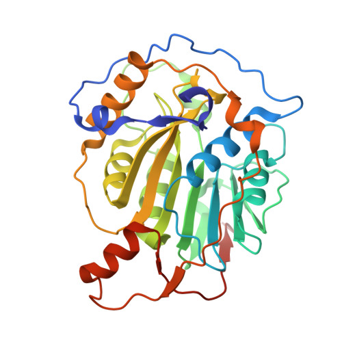
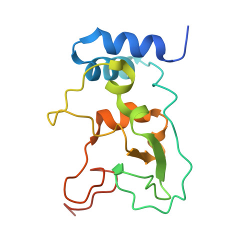
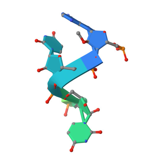
    2'-O-ribose methylation of the first transcribed base (adenine or A 1 in SARS-CoV-2) of viral RNA mimics host RNAs and subverts the innate immune response. How nsp16, with partner nsp10, assembles on the 5'-end of SARS-CoV-2 mRNA to methylate A 1 is not fully understood. We present a ∼2.4 Å crystal structure of the heterotetrameric complex formed by the cooperative assembly of two nsp16/nsp10 heterodimers with one 10-mer Cap-1 RNA (product) bound to each. An aromatic zipper-like motif in nsp16 and the N-terminal regions of nsp10 and nsp16 orchestrate oligomeric assembly for efficient methylation. The front catalytic pocket of nsp16 stabilizes the upstream portion of the RNA while downstream RNA remains unresolved, likely due to flexibility. An inverted nsp16 dimer extends the positively charged surface for longer RNA to influence catalysis. Additionally, a non-specific nucleotide-binding pocket on the backside of nsp16 plays a critical role in catalysis, contributing to enzymatic activity.


  • Organizational Affiliation
    • Greehey Children's Cancer Research Institute, University of Texas Health Science Center, San Antonio, TX 78229, USA; Department of Biochemistry and Structural Biology, University of Texas Health Science Center, San Antonio, TX 78229, USA.

Macromolecules

Find similar proteins by:  (by identity cutoff)  |  3D Structure
Entity ID: 1
MoleculeChains Sequence LengthOrganismDetailsImage
2'-O-methyltransferase
A, C
298Severe acute respiratory syndrome coronavirus 2Mutation(s): 0 
Gene Names: rep1a-1b
EC: 2.1.1
UniProt
Find proteins for P0DTD1 (Severe acute respiratory syndrome coronavirus 2)
Explore P0DTD1 
Go to UniProtKB:  P0DTD1
Entity Groups  
Sequence Clusters30% Identity50% Identity70% Identity90% Identity95% Identity100% Identity
UniProt GroupP0DTD1
Sequence Annotations
Expand
  • Reference Sequence
Find similar proteins by:  (by identity cutoff)  |  3D Structure
Entity ID: 2
MoleculeChains Sequence LengthOrganismDetailsImage
Non-structural protein 10
B, D
139Severe acute respiratory syndrome coronavirus 2Mutation(s): 0 
Gene Names: rep1a-1b
UniProt
Find proteins for P0DTD1 (Severe acute respiratory syndrome coronavirus 2)
Explore P0DTD1 
Go to UniProtKB:  P0DTD1
Entity Groups  
Sequence Clusters30% Identity50% Identity70% Identity90% Identity95% Identity100% Identity
UniProt GroupP0DTD1
Sequence Annotations
Expand
  • Reference Sequence

Find similar nucleic acids by:  Sequence   |   3D Structure  

Entity ID: 3
MoleculeChains LengthOrganismImage
RNA (5'-R(*AP*UP*UP*AP*AP*AP*GP*GP*UP*U)-3')
E, F
10Severe acute respiratory syndrome coronavirus 2
Sequence Annotations
Expand
  • Reference Sequence
Small Molecules
Ligands 5 Unique
IDChains Name / Formula / InChI Key2D Diagram3D Interactions
M7G
Query on M7G

Download Ideal Coordinates CCD File 
EA [auth E],
FA [auth F]
7N-METHYL-8-HYDROGUANOSINE-5'-DIPHOSPHATE
C11 H18 N5 O11 P2
SBASPRRECYVBRF-KQYNXXCUSA-O
SAH (Subject of Investigation/LOI)
Query on SAH

Download Ideal Coordinates CCD File 
H [auth A],
U [auth C]
S-ADENOSYL-L-HOMOCYSTEINE
C14 H20 N6 O5 S
ZJUKTBDSGOFHSH-WFMPWKQPSA-N
ZN
Query on ZN

Download Ideal Coordinates CCD File 
BA [auth D],
CA [auth D],
N [auth B],
O [auth B]
ZINC ION
Zn
PTFCDOFLOPIGGS-UHFFFAOYSA-N
EDO
Query on EDO

Download Ideal Coordinates CCD File 
AA [auth C]
DA [auth D]
I [auth A]
J [auth A]
K [auth A]
AA [auth C],
DA [auth D],
I [auth A],
J [auth A],
K [auth A],
L [auth A],
M [auth A],
P [auth B],
Q [auth B],
R [auth B],
S [auth B],
V [auth C],
W [auth C],
X [auth C],
Y [auth C],
Z [auth C]
1,2-ETHANEDIOL
C2 H6 O2
LYCAIKOWRPUZTN-UHFFFAOYSA-N
MG
Query on MG

Download Ideal Coordinates CCD File 
G [auth A],
T [auth C]
MAGNESIUM ION
Mg
JLVVSXFLKOJNIY-UHFFFAOYSA-N
Experimental Data & Validation

Experimental Data

  • Method: X-RAY DIFFRACTION
  • Resolution: 2.39 Å
  • R-Value Free:  0.219 (Depositor), 0.221 (DCC) 
  • R-Value Work:  0.180 (Depositor), 0.187 (DCC) 
  • R-Value Observed: 0.182 (Depositor) 
Space Group: P 1 21 1
Unit Cell:
Length ( Å )Angle ( ˚ )
a = 111.747α = 90
b = 58.893β = 95.481
c = 117.021γ = 90
Software Package:
Software NamePurpose
PHENIXrefinement
XDSdata reduction
Aimlessdata scaling
PHASERphasing

Structure Validation

View Full Validation Report


Ligand Structure Quality Assessment 


Entry History & Funding Information

Deposition Data


Funding OrganizationLocationGrant Number
National Institutes of Health/National Institute Of Allergy and Infectious Diseases (NIH/NIAID)United StatesR01AI161363

Revision History  (Full details and data files)

  • Version 1.0: 2025-02-05
    Type: Initial release
  • Version 1.1: 2026-02-18
    Changes: Database references