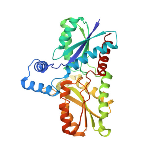
Entrance channels to coproheme in coproporphyrin ferrochelatase probed by exogenous imidazole binding.
Dali, A., Gabler, T., Sebastiani, F., Furtmuller, P.G., Becucci, M., Hofbauer, S., Smulevich, G.(2024) J Inorg Biochem 260: 112681-112681
- PubMed: 39146673
- DOI: https://doi.org/10.1016/j.jinorgbio.2024.112681
- Primary Citation of Related Structures:
9F0F, 9F0G - PubMed Abstract:
Iron insertion into porphyrins is an essential step in heme biosynthesis. In the coproporphyrin-dependent pathway, specific to monoderm bacteria, this reaction is catalyzed by the monomeric enzyme coproporphyrin ferrochelatase. In addition to the mechanistic details of the metalation of the porphyrin, the identification of the substrate access channel for ferrous iron to the active site is important to fully understand this enzymatic system. In fact, whether the iron reaches the active site from the distal or the proximal porphyrin side is still under debate. In this study we have thoroughly addressed this question in Listeria monocytogenes coproporphyrin ferrochelatase by X-ray crystallography, steady-state and pre-steady-state imidazole ligand binding studies, together with a detailed spectroscopic characterization using resonance Raman and UV-vis absorption spectroscopies in solution. Analysis of the X-ray structures of coproporphyrin ferrochelatase-coproporphyrin III crystals soaked with ferrous iron shows that iron is present on both sides of the porphyrin. The kinetic and spectroscopic study of imidazole binding to coproporphyrin ferrochelatase‑iron coproporphyrin III clearly indicates the presence of two possible binding sites in this monomeric enzyme that influence each other, which is confirmed by the observed cooperativity at steady-state and a biphasic behavior in the pre-steady-state experiments. The current results are discussed in the context of the entire heme biosynthetic pathway and pave the way for future studies focusing on protein-protein interactions.
- Dipartimento di Chimica "Ugo Schiff" (DICUS), Università di Firenze, Via della Lastruccia 3-13, I-50019 Sesto Fiorentino (FI), Italy.
Organizational Affiliation:


