9JUS | pdb_00009jus

Structure of villin bound to an actin trimer


Experimental Data Snapshot

  • Method: X-RAY DIFFRACTION
  • Resolution: 2.70 Å
  • R-Value Free: 
    0.226 (Depositor), 0.228 (DCC) 
  • R-Value Work: 
    0.196 (Depositor), 0.197 (DCC) 

Starting Models: experimental
View more details

wwPDB Validation   3D Report Full Report


Ligand Structure Quality Assessment 


This is version 1.1 of the entry. See complete history


Literature

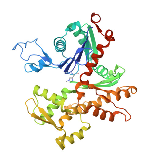
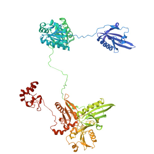
The structure of an actin nucleus stabilized by villin.

Robinson, R.C.Chongrungreang, T.Ponlachantra, K.Boonyarit, B.Dilly, G.F.Li, Y.I.Girguis, P.R.Copley, R.R.Claridge-Chang, A.

(2025) Sci Adv 11: eadw6915-eadw6915

  • DOI: https://doi.org/10.1126/sciadv.adw6915
  • Primary Citation of Related Structures:  
    9JUS, 9JVT, 9JW0

  • PubMed Abstract: 

    Villin is an actin filament nucleating, severing, capping and bundling protein; however, the structural basis for villin's functions and the characteristics of the actin polymerization nucleus remain poorly understood. Here, we present the structure of vent-worm villin bound to a trimeric actin nucleus. Villin wraps around and caps the barbed end of the actin trimer. Its headpiece domain interacts at the junction of two laterally associated actin protomers, leaving the pointed-end subunits open for elongation. Within the actin trimer, the two longitudinally associated subunits adopt barbed and pointed-end subunit conformations, while the lateral protomer exhibits a monomeric conformation. This provides the first view of an actin-filament nucleus, revealing that the transition into the filamentous form is stimulated and stabilized by the interactions with the pointed-end subunits. Our results also illuminate mechanisms of actin-filament dynamics and villin capping and severing, suggesting that F-to-G actin conformational transitions facilitate the later process.


  • Organizational Affiliation
    • School of Biomolecular Science and Engineering (BSE), Vidyasirimedhi Institute of Science and Technology (VISTEC), Rayong 21210 Thailand.

Macromolecules
Find similar proteins by:  (by identity cutoff)  |  3D Structure
Entity ID: 1
MoleculeChains Sequence LengthOrganismDetailsImage
Actin, alpha skeletal muscle377Gallus gallusMutation(s): 0 
Gene Names: ACTA1ACTA
EC: 3.6.4
UniProt
Find proteins for P68139 (Gallus gallus)
Explore P68139 
Go to UniProtKB:  P68139
Entity Groups  
Sequence Clusters30% Identity50% Identity70% Identity90% Identity95% Identity100% Identity
UniProt GroupP68139
Sequence Annotations
Expand
  • Reference Sequence
Find similar proteins by:  (by identity cutoff)  |  3D Structure
Entity ID: 2
MoleculeChains Sequence LengthOrganismDetailsImage
villinD [auth v],
H [auth V]
823Paralvinella sulfincolaMutation(s): 0 
UniProt
Find proteins for A0AAD9N3N6 (Paralvinella palmiformis)
Explore A0AAD9N3N6 
Go to UniProtKB:  A0AAD9N3N6
Entity Groups  
Sequence Clusters30% Identity50% Identity70% Identity90% Identity95% Identity100% Identity
UniProt GroupA0AAD9N3N6
Sequence Annotations
Expand
  • Reference Sequence
Small Molecules
Ligands 5 Unique
IDChains Name / Formula / InChI Key2D Diagram3D Interactions
ADP (Subject of Investigation/LOI)
Query on ADP

Download Ideal Coordinates CCD File 
BA [auth G]
I [auth p]
K [auth f]
M [auth g]
X [auth P]
BA [auth G],
I [auth p],
K [auth f],
M [auth g],
X [auth P],
Z [auth F]
ADENOSINE-5'-DIPHOSPHATE
C10 H15 N5 O10 P2
XTWYTFMLZFPYCI-KQYNXXCUSA-N
SCN
Query on SCN

Download Ideal Coordinates CCD File 
LA [auth V]THIOCYANATE ION
C N S
ZMZDMBWJUHKJPS-UHFFFAOYSA-M
CA (Subject of Investigation/LOI)
Query on CA

Download Ideal Coordinates CCD File 
DA [auth V]
EA [auth V]
FA [auth V]
GA [auth V]
HA [auth V]
DA [auth V],
EA [auth V],
FA [auth V],
GA [auth V],
HA [auth V],
IA [auth V],
JA [auth V],
KA [auth V],
O [auth v],
P [auth v],
Q [auth v],
R [auth v],
S [auth v],
T [auth v],
U [auth v],
V [auth v]
CALCIUM ION
Ca
BHPQYMZQTOCNFJ-UHFFFAOYSA-N
CL
Query on CL

Download Ideal Coordinates CCD File 
W [auth v]CHLORIDE ION
Cl
VEXZGXHMUGYJMC-UHFFFAOYSA-M
MG (Subject of Investigation/LOI)
Query on MG

Download Ideal Coordinates CCD File 
AA [auth F]
CA [auth G]
J [auth p]
L [auth f]
N [auth g]
AA [auth F],
CA [auth G],
J [auth p],
L [auth f],
N [auth g],
Y [auth P]
MAGNESIUM ION
Mg
JLVVSXFLKOJNIY-UHFFFAOYSA-N
Modified Residues  1 Unique
IDChains TypeFormula2D DiagramParent
HIC
Query on HIC
A [auth p]
B [auth f]
C [auth g]
E [auth P]
F
A [auth p],
B [auth f],
C [auth g],
E [auth P],
F,
G
L-PEPTIDE LINKINGC7 H11 N3 O2HIS
Experimental Data & Validation

Experimental Data

  • Method: X-RAY DIFFRACTION
  • Resolution: 2.70 Å
  • R-Value Free:  0.226 (Depositor), 0.228 (DCC) 
  • R-Value Work:  0.196 (Depositor), 0.197 (DCC) 
Space Group: P 1
Unit Cell:
Length ( Å )Angle ( ˚ )
a = 98.137α = 77.95
b = 102.432β = 72.59
c = 147.128γ = 65.96
Software Package:
Software NamePurpose
PHENIXrefinement
XDSdata reduction
Aimlessdata scaling
PHASERphasing

Structure Validation

View Full Validation Report


Ligand Structure Quality Assessment 


Entry History & Funding Information

Deposition Data


Funding OrganizationLocationGrant Number
Other privateMoore-Simons GBMF9743
Human Frontier Science Program (HFSP)FranceRGP0028/2018

Revision History  (Full details and data files)

  • Version 1.0: 2025-10-08
    Type: Initial release
  • Version 1.1: 2025-12-17
    Changes: Database references