


Crystal structure of H1 Haemagglutinin HN/18-HA2-L113S from Influenza A virus
Deng, G., Wei, X., Sun, H.To be published.
Experimental Data Snapshot
Starting Model: experimental
View more details
Entity ID: 1 | |||||
|---|---|---|---|---|---|
| Molecule | Chains | Sequence Length | Organism | Details | Image |
| Hemagglutinin | 321 | Influenza A virus | Mutation(s): 0 Gene Names: HA | 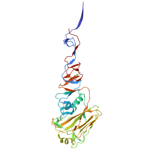 | |
UniProt | |||||
Find proteins for A0A6G5UYK1 (Influenza A virus) Explore A0A6G5UYK1 Go to UniProtKB: A0A6G5UYK1 | |||||
Entity Groups | |||||
| Sequence Clusters | 30% Identity50% Identity70% Identity90% Identity95% Identity100% Identity | ||||
| UniProt Group | A0A6G5UYK1 | ||||
Glycosylation | |||||
| Glycosylation Sites: 3 | |||||
Sequence AnnotationsExpand | |||||
| |||||
Entity ID: 2 | |||||
|---|---|---|---|---|---|
| Molecule | Chains | Sequence Length | Organism | Details | Image |
| Hemagglutinin | 156 | Influenza A virus | Mutation(s): 1 Gene Names: HA | 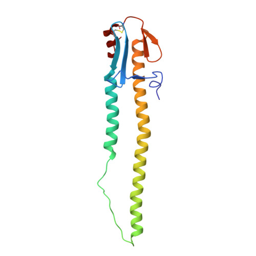 | |
UniProt | |||||
Find proteins for A0A6G5UYP3 (Influenza A virus) Explore A0A6G5UYP3 Go to UniProtKB: A0A6G5UYP3 | |||||
Entity Groups | |||||
| Sequence Clusters | 30% Identity50% Identity70% Identity90% Identity95% Identity100% Identity | ||||
| UniProt Group | A0A6G5UYP3 | ||||
Sequence AnnotationsExpand | |||||
| |||||
| Ligands 1 Unique | |||||
|---|---|---|---|---|---|
| ID | Chains | Name / Formula / InChI Key | 2D Diagram | 3D Interactions | |
| NAG Query on NAG | G [auth A] H [auth A] I [auth A] J [auth A] K [auth C] | 2-acetamido-2-deoxy-beta-D-glucopyranose C8 H15 N O6 OVRNDRQMDRJTHS-FMDGEEDCSA-N |  | ||
| Length ( Å ) | Angle ( ˚ ) |
|---|---|
| a = 65.091 | α = 90 |
| b = 249.627 | β = 118.43 |
| c = 65.509 | γ = 90 |
| Software Name | Purpose |
|---|---|
| REFMAC | refinement |
| HKL-3000 | data reduction |
| HKL-3000 | data scaling |
| PHENIX | phasing |
| Funding Organization | Location | Grant Number |
|---|---|---|
| Not funded | -- |


