9R08 | pdb_00009r08

Structure of the human heterotetrameric cis-prenyltransferase complex harboring NgBR-S249A in complex with magnesium, FsPP and IPP

  • Classification: TRANSFERASE
  • Organism(s): Homo sapiens
  • Expression System: Escherichia coli
  • Mutation(s): Yes 

  • Deposited: 2025-04-24 Released: 2025-10-29 
  • Deposition Author(s): Giladi, M., Haitin, Y.
  • Funding Organization(s): Israel Science Foundation, United States - Israel Binational Science Foundation (BSF), Other private

Experimental Data Snapshot

  • Method: X-RAY DIFFRACTION
  • Resolution: 2.40 Å
  • R-Value Free: 
    0.241 (Depositor), 0.241 (DCC) 
  • R-Value Work: 
    0.196 (Depositor), 0.197 (DCC) 
  • R-Value Observed: 
    0.198 (Depositor) 

Starting Model: experimental
View more details

wwPDB Validation   3D Report Full Report


Ligand Structure Quality Assessment 


This is version 1.1 of the entry. See complete history


Literature

Structural mechanisms of allosteric regulation in the human cis-prenyltransferase complex.

Giladi, M.Kredi, S.Guardiani, C.Aviram, L.Vankova, P.Gaizinger, Y.Man, P.Giacomello, A.Haitin, Y.

(2025) Nat Commun 16: 10786-10786

  • DOI: https://doi.org/10.1038/s41467-025-65833-6
  • Primary Citation of Related Structures:  
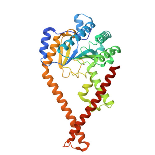
    9R08, 9R0E, 9R0K

  • PubMed Abstract: 

    Human cis-prenyltransferase (hcis-PT) synthesizes long-chain isoprenoids essential for N-linked protein glycosylation. This heteromeric complex comprises the catalytic subunit DHDDS and the regulatory Nogo-B receptor (NgBR). Although NgBR dramatically enhances DHDDS activity, the molecular basis for this allosteric regulation remains unclear. Here, we combined crystallography, hydrogen-deuterium exchange mass spectrometry (HDX-MS), molecular dynamics simulations, and network analysis to uncover the structural dynamics and communication pathways within hcis-PT. By solving the apo structure of hcis-PT, we reveal only a localized flexibility at the active site and the NgBR C-terminus. However, HDX-MS demonstrated widespread substrate-induced stabilization, particularly at the NgBR βD-βE loop, highlighting it as an allosteric hub. Functional mutagenesis scanning identified NgBR S249 as critical for enzymatic activity, independent of structural perturbations. Network analysis of MD simulations pinpointed this residue as a central node in inter-subunit communication, with perturbations disrupting downstream allosteric pathways, altering enzymatic activity. Our findings reveal a dynamic regulatory network centered at the inter-subunit interface, wherein specific NgBR residues modulate DHDDS activity through allosteric signaling. This work elucidates a conserved mechanism of subunit coordination in long-chain cis-prenyltransferases and suggests avenues for therapeutic targeting of hcis-PT-related disorders.


  • Organizational Affiliation
    • Department of Physiology and Pharmacology, Gray Faculty of Medical and Health Sciences, Tel-Aviv University, Tel-Aviv, Israel. moshegil@post.tau.ac.il.

Macromolecules
Find similar proteins by:  (by identity cutoff)  |  3D Structure
Entity ID: 1
MoleculeChains Sequence LengthOrganismDetailsImage
Dehydrodolichyl diphosphate synthase complex subunit DHDDS340Homo sapiensMutation(s): 0 
Gene Names: DHDDSHDS
EC: 2.5.1.87
UniProt & NIH Common Fund Data Resources
Find proteins for Q86SQ9 (Homo sapiens)
Explore Q86SQ9 
Go to UniProtKB:  Q86SQ9
PHAROS:  Q86SQ9
GTEx:  ENSG00000117682 
Entity Groups  
Sequence Clusters30% Identity50% Identity70% Identity90% Identity95% Identity100% Identity
UniProt GroupQ86SQ9
Sequence Annotations
Expand
  • Reference Sequence
Find similar proteins by:  (by identity cutoff)  |  3D Structure
Entity ID: 2
MoleculeChains Sequence LengthOrganismDetailsImage
Dehydrodolichyl diphosphate synthase complex subunit NUS1222Homo sapiensMutation(s): 1 
Gene Names: NUS1C6orf68NGBR
EC: 2.5.1.87
UniProt & NIH Common Fund Data Resources
Find proteins for Q96E22 (Homo sapiens)
Explore Q96E22 
Go to UniProtKB:  Q96E22
PHAROS:  Q96E22
GTEx:  ENSG00000153989 
Entity Groups  
Sequence Clusters30% Identity50% Identity70% Identity90% Identity95% Identity100% Identity
UniProt GroupQ96E22
Sequence Annotations
Expand
  • Reference Sequence
Experimental Data & Validation

Experimental Data

  • Method: X-RAY DIFFRACTION
  • Resolution: 2.40 Å
  • R-Value Free:  0.241 (Depositor), 0.241 (DCC) 
  • R-Value Work:  0.196 (Depositor), 0.197 (DCC) 
  • R-Value Observed: 0.198 (Depositor) 
Space Group: H 3 2
Unit Cell:
Length ( Å )Angle ( ˚ )
a = 184.399α = 90
b = 184.399β = 90
c = 112.575γ = 120
Software Package:
Software NamePurpose
PHENIXrefinement
PDB_EXTRACTdata extraction
XDSdata reduction
XDSdata scaling
PHASERphasing

Structure Validation

View Full Validation Report


Ligand Structure Quality Assessment 


Entry History & Funding Information

Deposition Data


Funding OrganizationLocationGrant Number
Israel Science FoundationIsrael1653/21
United States - Israel Binational Science Foundation (BSF)United States2023190
Other privateIsrael Cancer Research Fund 1289067

Revision History  (Full details and data files)

  • Version 1.0: 2025-10-29
    Type: Initial release
  • Version 1.1: 2025-12-10
    Changes: Database references