9SSA | pdb_00009ssa

Human angiotensin 1-converting enzyme C-domain in complex with zofenoprilat


Experimental Data Snapshot

  • Method: X-RAY DIFFRACTION
  • Resolution: 1.90 Å
  • R-Value Free: 
    0.218 (Depositor), 0.226 (DCC) 
  • R-Value Work: 
    0.181 (Depositor), 0.190 (DCC) 

Starting Model: experimental
View more details

wwPDB Validation   3D Report Full Report


Ligand Structure Quality Assessment 


This is version 1.0 of the entry. See complete history


Literature

Kinetic and structural characterisation of domain-specific angiotensin I-converting enzyme inhibition by captopril, rentiapril and zofenoprilat.

Gregory, K.S.Ramasamy, V.Sturrock, E.D.Acharya, K.R.

(2026) FEBS J 

  • DOI: https://doi.org/10.1111/febs.70428
  • Primary Citation of Related Structures:  
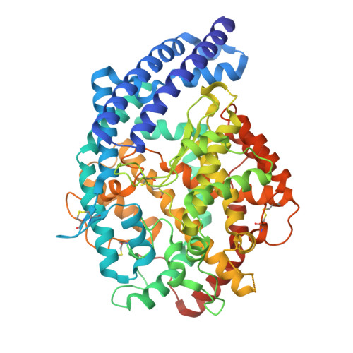
    9SS7, 9SS8, 9SS9, 9SSA, 9SSB

  • PubMed Abstract: 

    Angiotensin I-converting enzyme (ACE) is a zinc-dependent dipeptidyl carboxypeptidase involved in blood pressure regulation through proteolysis of angiotensin I (Ang-I) into the potent vasoconstrictor, angiotensin II (Ang-II). Inhibition of ACE is therefore used for the treatment of hypertension, heart failure, myocardial infarction, stroke and chronic kidney disease. Current ACE inhibitors (ACEi) bind both the N- and C-catalytic domains of ACE (referred to as nACE and cACE), and this has been linked to the occurrence of side effects due to the wide substrate specificity of ACE. The development of domain selective ACEi with reduced side effects is therefore key for improved therapeutic intervention. Understanding how current ACEi bind nACE and cACE, and their differences in domain selectivity should aid structure-based development of more selective ACEi by identifying different chemical groups that increase or decrease selectivity. We present the kinetic and structural characterisation of nACE and cACE with three thiolate ACEi, captopril (K i , nACE = 2.53 nm and cACE = 2.04 nm), rentiapril (monomer K i , nACE = 2.22 nm and cACE = 6.77 nm) and zofenoprilat (K i , nACE = 2.86 nm and cACE = 0.61 nm). Detailed structural analysis indicated that the S2' subsite likely contributes to the variation in domain selectivity observed for rentiapril and zofenoprilat due to differences in hydrophobicity and displacement of water molecules that contribute to ACE's hydration shell. Interestingly, in the cACE crystal structure, rentiapril bound as a dimer, and kinetic data revealed that both the monomeric and dimeric (dimer K i , nACE = 15.11 nm and cACE = 36.38 nm) forms of rentiapril inhibit ACE with nanomolar affinity.


  • Organizational Affiliation
    • Department of Life Sciences, University of Bath, UK.

Macromolecules
Find similar proteins by:  (by identity cutoff)  |  3D Structure
Entity ID: 1
MoleculeChains Sequence LengthOrganismDetailsImage
Angiotensin-converting enzyme597Homo sapiensMutation(s): 5 
Gene Names: ACEDCPDCP1
EC: 3.2.1 (PDB Primary Data), 3.4.15.1 (PDB Primary Data)
UniProt & NIH Common Fund Data Resources
Find proteins for P12821 (Homo sapiens)
Explore P12821 
Go to UniProtKB:  P12821
PHAROS:  P12821
GTEx:  ENSG00000159640 
Entity Groups  
Sequence Clusters30% Identity50% Identity70% Identity90% Identity95% Identity100% Identity
UniProt GroupP12821
Glycosylation
Glycosylation Sites: 2Go to GlyGen: P12821-1
Sequence Annotations
Expand
  • Reference Sequence
Oligosaccharides

Help

Entity ID: 2
MoleculeChains Length2D Diagram Glycosylation3D Interactions
alpha-D-mannopyranose-(1-3)-beta-D-mannopyranose-(1-4)-2-acetamido-2-deoxy-beta-D-glucopyranose-(1-4)-[alpha-L-fucopyranose-(1-6)]2-acetamido-2-deoxy-beta-D-glucopyranose
B
5N-Glycosylation
Small Molecules
Ligands 9 Unique
IDChains Name / Formula / InChI Key2D Diagram3D Interactions
ZED (Subject of Investigation/LOI)
Query on ZED

Download Ideal Coordinates CCD File 
E [auth A]L-PROLINE, 1-[(2S)-3-MERCAPTO-2-METHYL-1-OXOPROPYL]-4-(PHENYLTHIO)-, 4S
C15 H19 N O3 S2
UQWLOWFDKAFKAP-WXHSDQCUSA-N
NAG
Query on NAG

Download Ideal Coordinates CCD File 
C [auth A],
Q [auth A]
2-acetamido-2-deoxy-beta-D-glucopyranose
C8 H15 N O6
OVRNDRQMDRJTHS-FMDGEEDCSA-N
PEG
Query on PEG

Download Ideal Coordinates CCD File 
F [auth A],
H [auth A]
DI(HYDROXYETHYL)ETHER
C4 H10 O3
MTHSVFCYNBDYFN-UHFFFAOYSA-N
GOL
Query on GOL

Download Ideal Coordinates CCD File 
L [auth A]GLYCEROL
C3 H8 O3
PEDCQBHIVMGVHV-UHFFFAOYSA-N
IMD
Query on IMD

Download Ideal Coordinates CCD File 
P [auth A]IMIDAZOLE
C3 H5 N2
RAXXELZNTBOGNW-UHFFFAOYSA-O
ZN (Subject of Investigation/LOI)
Query on ZN

Download Ideal Coordinates CCD File 
D [auth A]ZINC ION
Zn
PTFCDOFLOPIGGS-UHFFFAOYSA-N
EDO
Query on EDO

Download Ideal Coordinates CCD File 
G [auth A],
I [auth A],
J [auth A],
K [auth A]
1,2-ETHANEDIOL
C2 H6 O2
LYCAIKOWRPUZTN-UHFFFAOYSA-N
BO3
Query on BO3

Download Ideal Coordinates CCD File 
M [auth A]BORIC ACID
B H3 O3
KGBXLFKZBHKPEV-UHFFFAOYSA-N
CL
Query on CL

Download Ideal Coordinates CCD File 
N [auth A],
O [auth A]
CHLORIDE ION
Cl
VEXZGXHMUGYJMC-UHFFFAOYSA-M
Modified Residues  1 Unique
IDChains TypeFormula2D DiagramParent
CSO
Query on CSO
A
L-PEPTIDE LINKINGC3 H7 N O3 SCYS
Experimental Data & Validation

Experimental Data

  • Method: X-RAY DIFFRACTION
  • Resolution: 1.90 Å
  • R-Value Free:  0.218 (Depositor), 0.226 (DCC) 
  • R-Value Work:  0.181 (Depositor), 0.190 (DCC) 
Space Group: P 21 21 21
Unit Cell:
Length ( Å )Angle ( ˚ )
a = 56.685α = 90
b = 85.007β = 90
c = 133.939γ = 90
Software Package:
Software NamePurpose
REFMACrefinement
DIALSdata reduction
Aimlessdata scaling
PHASERphasing

Structure Validation

View Full Validation Report


Ligand Structure Quality Assessment 


Entry History & Funding Information

Deposition Data


Funding OrganizationLocationGrant Number
Biotechnology and Biological Sciences Research Council (BBSRC)United KingdomBB/X001032/1

Revision History  (Full details and data files)

  • Version 1.0: 2026-02-18
    Type: Initial release