2GV0 | pdb_00002gv0

The structure of the orthorhombic form of soft-shelled turtle lysozyme at 1.9 angstroms resolution


Experimental Data Snapshot

  • Method: X-RAY DIFFRACTION
  • Resolution: 1.90 Å
  • R-Value Free: 
    0.232 (Depositor), 0.230 (DCC) 
  • R-Value Work: 
    0.207 (Depositor), 0.200 (DCC) 

wwPDB Validation   3D Report Full Report


This is version 1.4 of the entry. See complete history


Literature

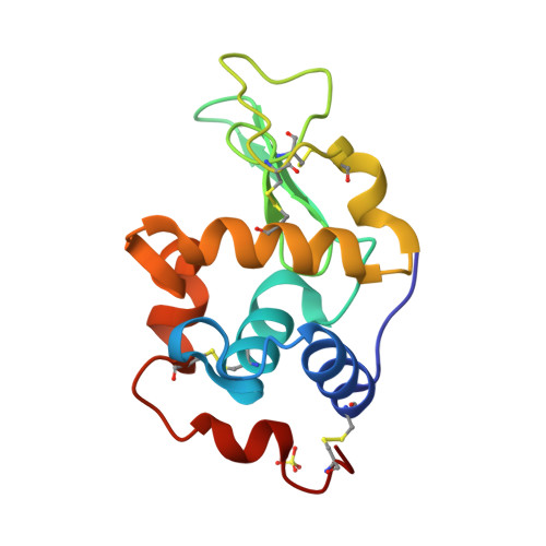
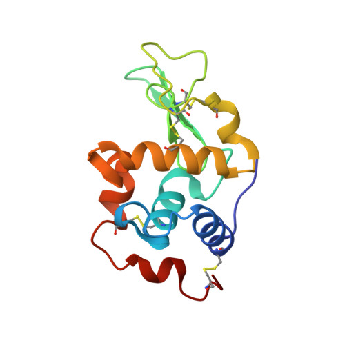
The 1.9 A X-ray structure of egg-white lysozyme from Taiwanese soft-shelled turtle (Trionyx Sinensis Wiegmann) exhibits structural differences from the standard chicken-type lysozyme.

Siritapetawee, J.Thammasirirak, S.Robinson, R.C.Yuvaniyama, J.

(2009) J Biochem 145: 193-198

  • DOI: https://doi.org/10.1093/jb/mvn156
  • Primary Citation of Related Structures:  
    2GV0

  • PubMed Abstract: 

    Lysozyme from Taiwanese soft-shelled turtle (SSTLB) has been purified from turtle egg white and crystallized. The crystals diffract X-rays beyond 2 A resolution and belong to the orthorhombic P2(1)2(1)2(1) space group containing one molecule per asymmetric unit. The structure was elucidated using pheasant egg-white lysozyme as the molecular replacement search template. The overall structure of SSTLB is very similar to that of hen egg-white lysozyme (HEWL). Nevertheless, Pro104 in the substrate subsite A and other amino acids in the substrate subsites E and F differ from those of HEWL. These substitutions result in local conformational differences in the substrate binding sites of the two enzymes, effecting substrate binding and transglycosylation efficiency, translating into differing ranges of products.


  • Organizational Affiliation

    College of Medicine and Public Health, Ubon Rajathanee University, Ubon Ratchathani, Thailand.


Macromolecules
Find similar proteins by:  (by identity cutoff)  |  3D Structure
Entity ID: 1
MoleculeChains Sequence LengthOrganismDetailsImage
Lysozyme C131Pelodiscus sinensisMutation(s): 0 
EC: 3.2.1.17
UniProt
Find proteins for Q7LZQ1 (Pelodiscus sinensis)
Explore Q7LZQ1 
Go to UniProtKB:  Q7LZQ1
Entity Groups  
Sequence Clusters30% Identity50% Identity70% Identity90% Identity95% Identity100% Identity
UniProt GroupQ7LZQ1
Sequence Annotations
Expand
  • Reference Sequence
Small Molecules
Ligands 1 Unique
IDChains Name / Formula / InChI Key2D Diagram3D Interactions
SO4
Query on SO4

Download Ideal Coordinates CCD File 
B [auth A]SULFATE ION
O4 S
QAOWNCQODCNURD-UHFFFAOYSA-L
Experimental Data & Validation

Experimental Data

  • Method: X-RAY DIFFRACTION
  • Resolution: 1.90 Å
  • R-Value Free:  0.232 (Depositor), 0.230 (DCC) 
  • R-Value Work:  0.207 (Depositor), 0.200 (DCC) 
Space Group: P 21 21 21
Unit Cell:
Length ( Å )Angle ( ˚ )
a = 37.758α = 90
b = 55.561β = 90
c = 72.234γ = 90
Software Package:
Software NamePurpose
d*TREKdata scaling
AMoREphasing
REFMACrefinement
PDB_EXTRACTdata extraction
CrystalCleardata reduction

Structure Validation

View Full Validation Report


Entry History 

Deposition Data

Revision History  (Full details and data files)

  • Version 1.0: 2007-05-08
    Type: Initial release
  • Version 1.1: 2008-05-01
    Changes: Version format compliance
  • Version 1.2: 2011-07-13
    Changes: Version format compliance
  • Version 1.3: 2024-11-20
    Changes: Data collection, Database references, Derived calculations, Structure summary
  • Version 1.4: 2025-01-29
    Changes: Database references