7EE8 | pdb_00007ee8

The crystal structure of MIF bound to compound D5


Experimental Data Snapshot

  • Method: X-RAY DIFFRACTION
  • Resolution: 1.22 Å
  • R-Value Free: 
    0.242 (Depositor), 0.241 (DCC) 
  • R-Value Work: 
    0.214 (Depositor), 0.213 (DCC) 
  • R-Value Observed: 
    0.214 (Depositor) 

wwPDB Validation   3D Report Full Report


Ligand Structure Quality Assessment 

Created with Raphaël 2.3.0Worse 01 BetterLigand structure goodness of fit to experimental dataBest fitted 7Y8Click on this verticalbar to view details

This is version 1.0 of the entry. See complete history


Literature

Repurposing old drugs as novel inhibitors of human MIF from structural and functional analysis.

Yang, L.Yang, C.Wang, L.Yang, Z.Guo, D.Fan, C.

(2022) Bioorg Med Chem Lett 55: 128445-128445

  • DOI: https://doi.org/10.1016/j.bmcl.2021.128445
  • Primary Citation of Related Structures:  
    7EDQ, 7EE8

  • PubMed Abstract: 

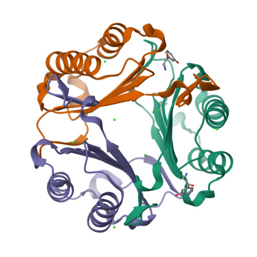
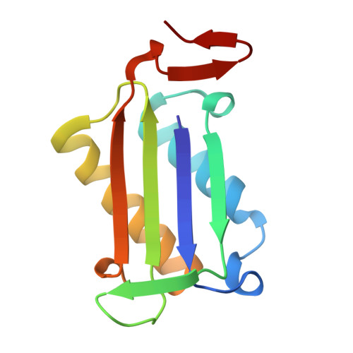
    Human macrophage migration inhibitory factor (MIF) is an important pro-inflammatory cytokine that plays multiple pleiotropic functions. It is considered as a promising therapeutic target for the infectious, autoimmune, and cardiovascular diseases and cancers. The development of MIF inhibitors has not been translated into clinical success despite decades of research. Given the time and cost of developing new drugs, existing drugs with clarified safety and pharmacokinetics are explored for their potential as novel MIF inhibitors. This study identified five known drugs that could inhibit MIF's tautomerase activity and MIF-mediated cell chemotaxis in RAW264.7 cells. It was found that compounds D2 (histamine), D5 (metaraminol), and D8 (nebivolol) exhibited micromolar-range inhibition potency close to the positive control ISO-1. Kinetics and the mechanism for inhibition were subsequently determined. Moreover, the detailed inhibitor-binding patterns were investigated by X-ray crystallography, computational molecular docking, and structure-based analysis. Therefore, this study elucidates the molecular mechanism of repurposed drugs acting on MIF and provides a structural foundation for lead optimization to promote the clinical development of MIF-targeted drugs.


  • Organizational Affiliation

    School of Basic Medical Sciences, Wuhan University, Wuhan 430071, PR China.


Macromolecules
Find similar proteins by:  (by identity cutoff)  |  3D Structure
Entity ID: 1
MoleculeChains Sequence LengthOrganismDetailsImage
Macrophage migration inhibitory factor
A, B, C
114Homo sapiensMutation(s): 0 
Gene Names: MIFGLIFMMIF
EC: 5.3.2.1 (PDB Primary Data), 5.3.3.12 (PDB Primary Data)
UniProt & NIH Common Fund Data Resources
Find proteins for P14174 (Homo sapiens)
Explore P14174 
Go to UniProtKB:  P14174
PHAROS:  P14174
GTEx:  ENSG00000240972 
Entity Groups  
Sequence Clusters30% Identity50% Identity70% Identity90% Identity95% Identity100% Identity
UniProt GroupP14174
Sequence Annotations
Expand
  • Reference Sequence
Small Molecules
Ligands 3 Unique
IDChains Name / Formula / InChI Key2D Diagram3D Interactions
J23
Query on J23

Download Ideal Coordinates CCD File 
K [auth B]3-[(2S)-2-azanyl-1-oxidanyl-propyl]phenol
C9 H13 N O2
WXFIGDLSSYIKKV-IMTBSYHQSA-N
7Y8 (Subject of Investigation/LOI)
Query on 7Y8

Download Ideal Coordinates CCD File 
G [auth A]3-[(2R)-2-azanyl-1-oxidanyl-propyl]phenol
C9 H13 N O2
WXFIGDLSSYIKKV-VJSCVCEBSA-N
CL
Query on CL

Download Ideal Coordinates CCD File 
D [auth A]
E [auth A]
F [auth A]
H [auth B]
I [auth B]
CHLORIDE ION
Cl
VEXZGXHMUGYJMC-UHFFFAOYSA-M
Experimental Data & Validation

Experimental Data

  • Method: X-RAY DIFFRACTION
  • Resolution: 1.22 Å
  • R-Value Free:  0.242 (Depositor), 0.241 (DCC) 
  • R-Value Work:  0.214 (Depositor), 0.213 (DCC) 
  • R-Value Observed: 0.214 (Depositor) 
Space Group: P 21 21 21
Unit Cell:
Length ( Å )Angle ( ˚ )
a = 67.775α = 90
b = 68.144β = 90
c = 86.162γ = 90
Software Package:
Software NamePurpose
PHENIXrefinement
PDB_EXTRACTdata extraction
HKL-2000data reduction
SCALEPACKdata scaling
PHENIXphasing

Structure Validation

View Full Validation Report


Ligand Structure Quality Assessment 

Created with Raphaël 2.3.0Worse 01 BetterLigand structure goodness of fit to experimental dataBest fitted 7Y8Click on this verticalbar to view details

Entry History & Funding Information

Deposition Data


Funding OrganizationLocationGrant Number
National Natural Science Foundation of China (NSFC)China31570762

Revision History  (Full details and data files)

  • Version 1.0: 2025-03-26
    Type: Initial release