9MXZ | pdb_00009mxz

Lecithin:Cholesterol Acyltransferase Bound to Apolipoprotein A-I dimer in HDL


Experimental Data Snapshot

  • Method: ELECTRON MICROSCOPY
  • Resolution: 9.80 Å
  • Aggregation State: PARTICLE 
  • Reconstruction Method: SINGLE PARTICLE 

wwPDB Validation   3D Report Full Report


This is version 1.1 of the entry. See complete history


Literature

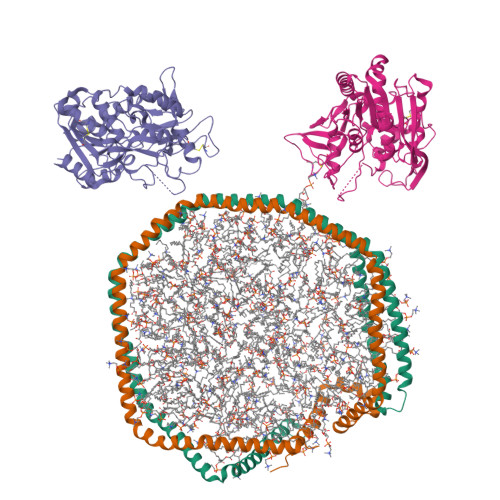
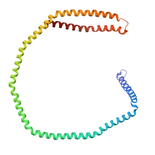
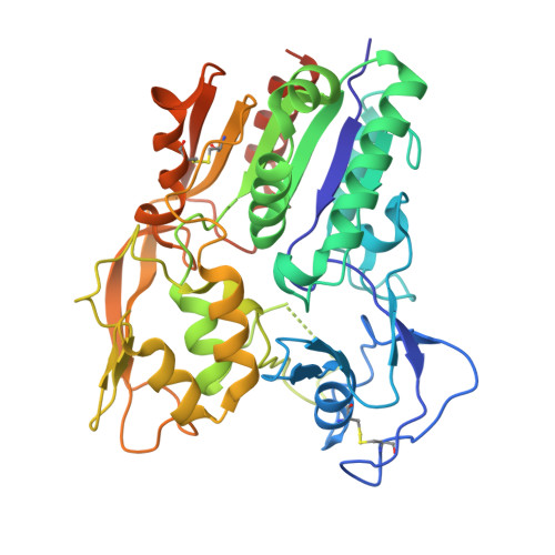
Lecithin:Cholesterol Acyltransferase Binds a Discontinuous Binding Site on Adjacent Apolipoprotein A-I Belts in HDL.

Coleman, B.Bedi, S.Hill, J.H.Morris, J.Manthei, K.A.Hart, R.C.He, Y.Shah, A.S.Jerome, W.G.Vaisar, T.Bornfeldt, K.E.Song, H.Segrest, J.P.Heinecke, J.W.Aller, S.G.Tesmer, J.J.G.Davidson, W.S.

(2025) J Lipid Res : 100786-100786

  • DOI: https://doi.org/10.1016/j.jlr.2025.100786
  • Primary Citation of Related Structures:  
    9MXZ

  • PubMed Abstract: 

    Lecithin:cholesterol acyltransferase (LCAT) is a high-density lipoprotein (HDL) modifying protein that profoundly affects the composition and function of HDL subspecies. The cholesterol esterification activity of LCAT is dramatically increased by apolipoprotein A-I (APOA1) on HDL, but the mechanism remains unclear. Using site-directed mutagenesis, cross-linking, mass spectrometry, electron microscopy, protein engineering and molecular docking, we identified two LCAT binding sites formed by helices 4 and 6 from two antiparallel APOA1 molecules in HDL. Although the reciprocating APOA1 'belts' form two ostensibly symmetrical binding locations, LCAT can adopt distinct orientations at each site, as shown by our 9.8 Å cryoEM envelope. In one case, LCAT membrane binding domains align with the APOA1 belts and, in the other, the HDL phospholipids. By introducing disulfide bonds between the APOA1 helical domains, we demonstrated that LCAT does not require helical separation during its reaction cycle. This indicates that LCAT, anchored to APOA1 belts, accesses substrates and deposits products through interactions with the planar lipid surface. This model of the LCAT/APOA1 interaction provides insights into how LCAT and possibly other HDL-modifying factors engage the APOA1 scaffold, offering potential strategies to enhance LCAT activity in individuals with genetic defects.


  • Organizational Affiliation

    Department of Molecular and Cellular Biosciences, University of Cincinnati, Cincinnati, OH, USA.


Macromolecules
Find similar proteins by:  (by identity cutoff)  |  3D Structure
Entity ID: 1
MoleculeChains Sequence LengthOrganismDetailsImage
Apolipoprotein A-IA [auth E],
B [auth A]
243Homo sapiensMutation(s): 0 
Gene Names: APOA1
UniProt & NIH Common Fund Data Resources
Find proteins for P02647 (Homo sapiens)
Explore P02647 
Go to UniProtKB:  P02647
PHAROS:  P02647
GTEx:  ENSG00000118137 
Entity Groups  
Sequence Clusters30% Identity50% Identity70% Identity90% Identity95% Identity100% Identity
UniProt GroupP02647
Sequence Annotations
Expand
  • Reference Sequence
Find similar proteins by:  (by identity cutoff)  |  3D Structure
Entity ID: 2
MoleculeChains Sequence LengthOrganismDetailsImage
Phosphatidylcholine-sterol acyltransferaseC [auth B],
D [auth C]
396Homo sapiensMutation(s): 0 
Gene Names: LCAT
EC: 2.3.1.43 (PDB Primary Data), 3.1.1.47 (PDB Primary Data)
UniProt & NIH Common Fund Data Resources
Find proteins for P04180 (Homo sapiens)
Explore P04180 
Go to UniProtKB:  P04180
PHAROS:  P04180
GTEx:  ENSG00000213398 
Entity Groups  
Sequence Clusters30% Identity50% Identity70% Identity90% Identity95% Identity100% Identity
UniProt GroupP04180
Sequence Annotations
Expand
  • Reference Sequence
Small Molecules
Ligands 1 Unique
IDChains Name / Formula / InChI Key2D Diagram3D Interactions
6PL
Query on 6PL

Download Ideal Coordinates CCD File 
AA [auth E]
AB [auth E]
AC [auth A]
AD [auth A]
AE [auth A]
(4S,7R)-4-HYDROXY-N,N,N-TRIMETHYL-9-OXO-7-[(PALMITOYLOXY)METHYL]-3,5,8-TRIOXA-4-PHOSPHAHEXACOSAN-1-AMINIUM 4-OXIDE
C42 H85 N O8 P
PZNPLUBHRSSFHT-RRHRGVEJSA-O
Experimental Data & Validation

Experimental Data

  • Method: ELECTRON MICROSCOPY
  • Resolution: 9.80 Å
  • Aggregation State: PARTICLE 
  • Reconstruction Method: SINGLE PARTICLE 

Structure Validation

View Full Validation Report


Entry History & Funding Information

Deposition Data


Funding OrganizationLocationGrant Number
National Institutes of Health/National Heart, Lung, and Blood Institute (NIH/NHLBI)United StatesP01 HL116263
National Institutes of Health/National Heart, Lung, and Blood Institute (NIH/NHLBI)United StatesR01 HL153118

Revision History  (Full details and data files)

  • Version 1.0: 2025-04-02
    Type: Initial release
  • Version 1.1: 2025-04-09
    Changes: Data collection, Database references