9QFU | pdb_00009qfu

Human Tryptase beta-2 (hTPSB2) complexed with covalent inhibitor Compound #1


Experimental Data Snapshot

  • Method: X-RAY DIFFRACTION
  • Resolution: 1.98 Å
  • R-Value Free: 
    0.221 (Depositor), 0.226 (DCC) 
  • R-Value Work: 
    0.177 (Depositor), 0.184 (DCC) 
  • R-Value Observed: 
    0.179 (Depositor) 

Starting Model: experimental
View more details

wwPDB Validation   3D Report Full Report


Ligand Structure Quality Assessment 

Created with Raphaël 2.3.0Worse 01 BetterLigand structure goodness of fit to experimental dataBest fitted A1I52Click on this verticalbar to view detailsBest fitted PEGClick on this verticalbar to view detailsBest fitted SO4Click on this verticalbar to view detailsBest fitted GOLClick on this verticalbar to view detailsBest fitted EDOClick on this verticalbar to view detailsBest fitted ACTClick on this verticalbar to view details

This is version 1.1 of the entry. See complete history


Literature

Integrating Surface Plasmon Resonance and Docking Analysis for Mechanistic Insights of Tryptase Inhibitors.

Porta, A.Manelfi, C.Talarico, C.Beccari, A.R.Brindisi, M.Summa, V.Iaconis, D.Gobbi, M.Beeg, M.

(2025) Molecules 30

  • DOI: https://doi.org/10.3390/molecules30061338
  • Primary Citation of Related Structures:  
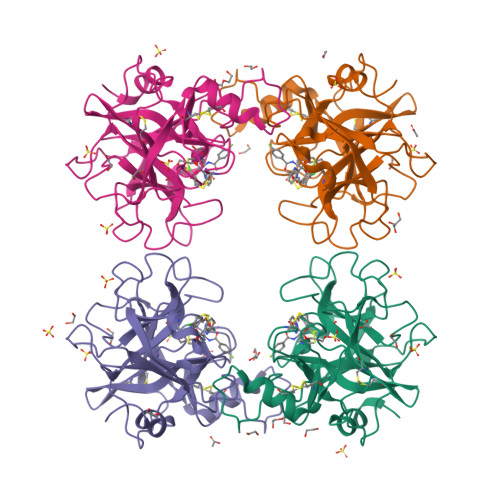
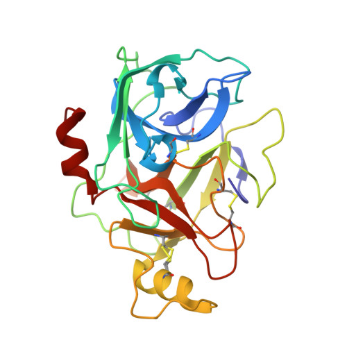
    9QFU, 9QFV

  • PubMed Abstract: 

    Tryptase is a tetrameric serine protease and a key component of mast cell granules. Here, we explored an integrated approach to characterize tryptase ligands, combining novel experimental binding studies using Surface Plasmon Resonance, with in silico analysis through the Exscalate platform. For this, we focused on three inhibitors previously reported in the literature, including a bivalent inhibitor and its corresponding monovalent compound. All three ligands showed concentration-dependent binding to immobilized human tryptase with the bivalent inhibitor showing the highest affinity. Furthermore, Rmax values were similar, indicating that the compounds occupy all four binding pockets of the tryptase tetramer. This hypothesis was supported by in silico computational analysis that revealed the binding mode of the monovalent ligand, one in each monomer pocket, compared with crystal structure of the bivalent one, which simultaneously occupies two binding pockets. Additionally, we solved the 2.06 Å X-ray crystal structures of human Tryptase beta-2 (hTPSB2), in both its apo form and in complex with compound # 1 , experimentally confirming the binding mode and the key molecular interactions predicted by docking studies for this compound. This integrated approach offers a robust framework for elucidating both the strength and mode of interaction of potential tryptase inhibitors.


  • Organizational Affiliation

    Istituto di Ricerche Farmacologiche Mario Negri IRCCS, Via Mario Negri, 2, 20156 Milano, Italy.


Macromolecules
Find similar proteins by:  (by identity cutoff)  |  3D Structure
Entity ID: 1
MoleculeChains Sequence LengthOrganismDetailsImage
Tryptase beta-2
A, B, C, D
243Homo sapiensMutation(s): 3 
EC: 3.4.21.59
UniProt & NIH Common Fund Data Resources
Find proteins for P20231 (Homo sapiens)
Explore P20231 
Go to UniProtKB:  P20231
PHAROS:  P20231
GTEx:  ENSG00000197253 
Entity Groups  
Sequence Clusters30% Identity50% Identity70% Identity90% Identity95% Identity100% Identity
UniProt GroupP20231
Sequence Annotations
Expand
  • Reference Sequence
Small Molecules
Ligands 6 Unique
IDChains Name / Formula / InChI Key2D Diagram3D Interactions
A1I52 (Subject of Investigation/LOI)
Query on A1I52

Download Ideal Coordinates CCD File 
E [auth A],
JA [auth D],
O [auth B],
X [auth C]
~{N}-[(1~{S},2~{S})-6-azanyl-1-[5-[[4-[2-(3,4-dichlorophenyl)ethoxy]phenyl]methyl]-1,2,4-oxadiazol-3-yl]-1-oxidanyl-hexan-2-yl]-4-fluoranyl-benzamide
C30 H31 Cl2 F N4 O4
VDZJQCQLVMCFBD-XCZPVHLTSA-N
PEG (Subject of Investigation/LOI)
Query on PEG

Download Ideal Coordinates CCD File 
J [auth A],
QA [auth D]
DI(HYDROXYETHYL)ETHER
C4 H10 O3
MTHSVFCYNBDYFN-UHFFFAOYSA-N
SO4 (Subject of Investigation/LOI)
Query on SO4

Download Ideal Coordinates CCD File 
AA [auth C]
BA [auth C]
EA [auth C]
F [auth A]
G [auth A]
SULFATE ION
O4 S
QAOWNCQODCNURD-UHFFFAOYSA-L
GOL (Subject of Investigation/LOI)
Query on GOL

Download Ideal Coordinates CCD File 
S [auth B]GLYCEROL
C3 H8 O3
PEDCQBHIVMGVHV-UHFFFAOYSA-N
EDO (Subject of Investigation/LOI)
Query on EDO

Download Ideal Coordinates CCD File 
CA [auth C]
FA [auth C]
GA [auth C]
HA [auth C]
IA [auth C]
1,2-ETHANEDIOL
C2 H6 O2
LYCAIKOWRPUZTN-UHFFFAOYSA-N
ACT (Subject of Investigation/LOI)
Query on ACT

Download Ideal Coordinates CCD File 
DA [auth C],
N [auth A],
R [auth B]
ACETATE ION
C2 H3 O2
QTBSBXVTEAMEQO-UHFFFAOYSA-M
Experimental Data & Validation

Experimental Data

  • Method: X-RAY DIFFRACTION
  • Resolution: 1.98 Å
  • R-Value Free:  0.221 (Depositor), 0.226 (DCC) 
  • R-Value Work:  0.177 (Depositor), 0.184 (DCC) 
  • R-Value Observed: 0.179 (Depositor) 
Space Group: P 31
Unit Cell:
Length ( Å )Angle ( ˚ )
a = 78.552α = 90
b = 78.552β = 90
c = 165.688γ = 120
Software Package:
Software NamePurpose
REFMACrefinement
autoPROCdata reduction
autoPROCdata scaling
REFMACphasing

Structure Validation

View Full Validation Report


Ligand Structure Quality Assessment 

Created with Raphaël 2.3.0Worse 01 BetterLigand structure goodness of fit to experimental dataBest fitted A1I52Click on this verticalbar to view detailsBest fitted PEGClick on this verticalbar to view detailsBest fitted SO4Click on this verticalbar to view detailsBest fitted GOLClick on this verticalbar to view detailsBest fitted EDOClick on this verticalbar to view detailsBest fitted ACTClick on this verticalbar to view details

Entry History & Funding Information

Deposition Data


Funding OrganizationLocationGrant Number
Not funded--

Revision History  (Full details and data files)

  • Version 1.0: 2025-03-26
    Type: Initial release
  • Version 1.1: 2025-04-09
    Changes: Database references