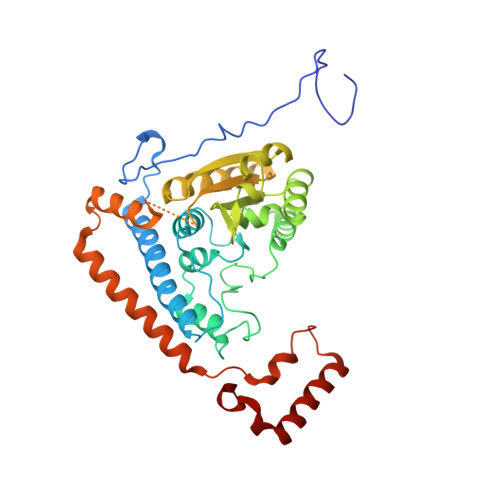
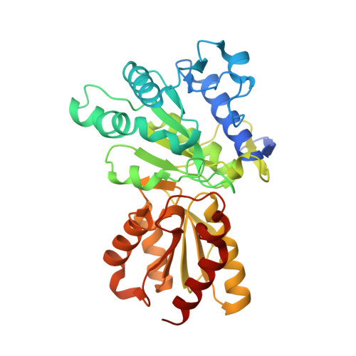
1X80

Crystal structure of the human mitochondrial branched-chain alpha-ketoacid dehydrogenase


Experimental Data Snapshot

  • Method: X-RAY DIFFRACTION
  • Resolution: 2.00 Å
  • R-Value Free: 0.193 
  • R-Value Work: 0.160 
  • R-Value Observed: 0.161 

Starting Model: experimental
View more details

wwPDB Validation   3D Report Full Report


Ligand Structure Quality Assessment 


This is version 2.2 of the entry. See complete history


Literature

Molecular mechanism for regulation of the human mitochondrial branched-chain alpha-ketoacid dehydrogenase complex by phosphorylation

Wynn, R.M.Kato, M.Machius, M.Chuang, J.L.Li, J.Tomchick, D.R.Chuang, D.T.

(2004) Structure 12: 2185-2196

  • DOI: https://doi.org/10.1016/j.str.2004.09.013
  • Primary Citation of Related Structures:  
    1U5B, 1X7W, 1X7X, 1X7Y, 1X7Z, 1X80

  • PubMed Abstract: 

    The human mitochondrial branched-chain alpha-ketoacid dehydrogenase complex (BCKDC) is a 4 MDa macromolecular machine comprising three catalytic components (E1b, E2b, and E3), a kinase, and a phosphatase. The BCKDC overall activity is tightly regulated by phosphorylation in response to hormonal and dietary stimuli. We report that phosphorylation of Ser292-alpha in the E1b active site channel results in an order-to-disorder transition of the conserved phosphorylation loop carrying the phosphoryl serine. The conformational change is triggered by steric clashes of the phosphoryl group with invariant His291-alpha that serves as an indispensable anchor for the phosphorylation loop through bound thiamin diphosphate. Phosphorylation of Ser292-alpha does not severely impede the E1b-dependent decarboxylation of alpha-ketoacids. However, the disordered loop conformation prevents phosphorylated E1b from binding the E2b lipoyl-bearing domain, which effectively shuts off the E1b-catalyzed reductive acylation reaction and therefore completely inactivates BCKDC. This mechanism provides a paradigm for regulation of mitochondrial alpha-ketoacid dehydrogenase complexes by phosphorylation.


  • Organizational Affiliation

    Department of Biochemistry, University of Texas Southwestern Medical Center, Dallas, TX 75390, USA.


Macromolecules
Find similar proteins by:  (by identity cutoff)  |  3D Structure
Entity ID: 1
MoleculeChains Sequence LengthOrganismDetailsImage
2-oxoisovalerate dehydrogenase alpha subunit400Homo sapiensMutation(s): 1 
Gene Names: BCKDHA
EC: 1.2.4.4
UniProt & NIH Common Fund Data Resources
Find proteins for P12694 (Homo sapiens)
Explore P12694 
Go to UniProtKB:  P12694
PHAROS:  P12694
GTEx:  ENSG00000248098 
Entity Groups  
Sequence Clusters30% Identity50% Identity70% Identity90% Identity95% Identity100% Identity
UniProt GroupP12694
Sequence Annotations
Expand
  • Reference Sequence
Find similar proteins by:  (by identity cutoff)  |  3D Structure
Entity ID: 2
MoleculeChains Sequence LengthOrganismDetailsImage
2-oxoisovalerate dehydrogenase beta subunit342Homo sapiensMutation(s): 0 
Gene Names: BCKDHB
EC: 1.2.4.4
UniProt & NIH Common Fund Data Resources
Find proteins for P21953 (Homo sapiens)
Explore P21953 
Go to UniProtKB:  P21953
PHAROS:  P21953
GTEx:  ENSG00000083123 
Entity Groups  
Sequence Clusters30% Identity50% Identity70% Identity90% Identity95% Identity100% Identity
UniProt GroupP21953
Sequence Annotations
Expand
  • Reference Sequence
Small Molecules
Ligands 5 Unique
IDChains Name / Formula / InChI Key2D Diagram3D Interactions
TPP
Query on TPP

Download Ideal Coordinates CCD File 
F [auth A]THIAMINE DIPHOSPHATE
C12 H19 N4 O7 P2 S
AYEKOFBPNLCAJY-UHFFFAOYSA-O
GOL
Query on GOL

Download Ideal Coordinates CCD File 
H [auth B]GLYCEROL
C3 H8 O3
PEDCQBHIVMGVHV-UHFFFAOYSA-N
MN
Query on MN

Download Ideal Coordinates CCD File 
D [auth A]MANGANESE (II) ION
Mn
WAEMQWOKJMHJLA-UHFFFAOYSA-N
K
Query on K

Download Ideal Coordinates CCD File 
C [auth A],
G [auth B]
POTASSIUM ION
K
NPYPAHLBTDXSSS-UHFFFAOYSA-N
CL
Query on CL

Download Ideal Coordinates CCD File 
E [auth A]CHLORIDE ION
Cl
VEXZGXHMUGYJMC-UHFFFAOYSA-M
Modified Residues  1 Unique
IDChains TypeFormula2D DiagramParent
SEP
Query on SEP
A
L-PEPTIDE LINKINGC3 H8 N O6 PSER
Experimental Data & Validation

Experimental Data

  • Method: X-RAY DIFFRACTION
  • Resolution: 2.00 Å
  • R-Value Free: 0.193 
  • R-Value Work: 0.160 
  • R-Value Observed: 0.161 
  • Space Group: P 31 2 1
Unit Cell:
Length ( Å )Angle ( ˚ )
a = 144.403α = 90
b = 144.403β = 90
c = 69.009γ = 120
Software Package:
Software NamePurpose
HKL-2000data collection
HKL-2000data reduction
AMoREphasing
REFMACrefinement
HKL-2000data scaling

Structure Validation

View Full Validation Report


Ligand Structure Quality Assessment 


Entry History 

Deposition Data

Revision History  (Full details and data files)

  • Version 1.0: 2004-11-23
    Type: Initial release
  • Version 1.1: 2008-04-30
    Changes: Version format compliance
  • Version 1.2: 2011-07-13
    Changes: Derived calculations, Version format compliance
  • Version 2.0: 2021-08-04
    Changes: Atomic model, Data collection, Database references, Derived calculations, Non-polymer description, Structure summary
  • Version 2.1: 2023-08-23
    Changes: Data collection, Database references, Refinement description
  • Version 2.2: 2024-10-30
    Changes: Structure summary