


Molecular Basis of c-MET Inhibition by Approved Small Molecule Drugs: A Structural Perspective
Russell, I.C., Bachurska-Szpala, P., van Beek, L., Michaelides, I.N., Phillips, C., Snijder, A., Stubbs, C.J., Collie, G.W.(2026) ACS Med Chem Lett
Experimental Data Snapshot
Starting Model: experimental
View more details
wwPDB Validation 3D Report Full Report

(2026) ACS Med Chem Lett
Entity ID: 1 | |||||
|---|---|---|---|---|---|
| Molecule | Chains | Sequence Length | Organism | Details | Image |
| Hepatocyte growth factor receptor | 296 | Homo sapiens | Mutation(s): 0 Gene Names: MET EC: 2.7.10.1 | 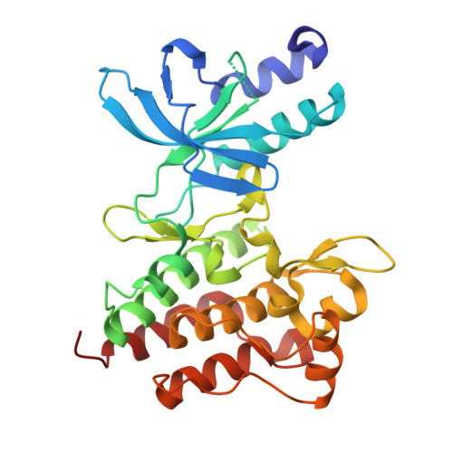 | |
UniProt & NIH Common Fund Data Resources | |||||
Find proteins for P08581 (Homo sapiens) Explore P08581 Go to UniProtKB: P08581 | |||||
PHAROS: P08581 GTEx: ENSG00000105976 | |||||
Entity Groups | |||||
| Sequence Clusters | 30% Identity50% Identity70% Identity90% Identity95% Identity100% Identity | ||||
| UniProt Group | P08581 | ||||
Sequence AnnotationsExpand | |||||
| |||||
| Ligands 3 Unique | |||||
|---|---|---|---|---|---|
| ID | Chains | Name / Formula / InChI Key | 2D Diagram | 3D Interactions | |
| 15P Query on 15P | C [auth A], E [auth A] | POLYETHYLENE GLYCOL (N=34) C69 H140 O35 VUYXVWGKCKTUMF-UHFFFAOYSA-N |  | ||
| A1JT6 (Subject of Investigation/LOI) Query on A1JT6 | B [auth A] | Glumetinib C21 H17 N9 O2 S RYBLECYFLJXEJX-UHFFFAOYSA-N |  | ||
| PEG Query on PEG | D [auth A] | DI(HYDROXYETHYL)ETHER C4 H10 O3 MTHSVFCYNBDYFN-UHFFFAOYSA-N |  | ||
| Length ( Å ) | Angle ( ˚ ) |
|---|---|
| a = 37.958 | α = 90 |
| b = 42.719 | β = 92.5 |
| c = 85.287 | γ = 90 |
| Software Name | Purpose |
|---|---|
| BUSTER | refinement |
| autoPROC | data reduction |
| autoPROC | data scaling |
| PHASER | phasing |
| Funding Organization | Location | Grant Number |
|---|---|---|
| Not funded | -- |


